| Journal of Clinical Gynecology and Obstetrics, ISSN 1927-1271 print, 1927-128X online, Open Access |
| Article copyright, the authors; Journal compilation copyright, J Clin Gynecol Obstet and Elmer Press Inc |
| Journal website https://jcgo.elmerpub.com |
Case Report
Volume 14, Number 4, December 2025, pages 184-188

Uterine Fibroid Embolization in Patient With Symptomatic Uterine Leiomyomas in Uterine Didelphys
Nicole T. Stamboa, c, Glenn W. Stambob
aLake Erie College of Osteopathic Medicine, Bradenton, FL 34211,
USA
bDivision of Vascular and Interventional Radiology, Department of Radiology,
AdventHealth Carrollwood Hospital Tampa, FL, USA
cCorresponding Author: Nicole
T. Stambo, Lake Erie College of Osteopathic Medicine, Bradenton, FL 34211, USA
Manuscript submitted August 12, 2025, accepted November 31, 2025, published online December 11,
2025
Short title: UFE in Uterine Didelphys With Leiomyomas
doi:
https://doi.org/10.14740/jcgo1532
| Abstract | ▴Top |
Uterine didelphys is a rare congenital anomaly resulting from compete failure of Mullerian duct fusion, occurring in 0.5-5% of women. Its coexistence with uterine leiomyomas is extremely uncommon, with an estimated prevalence of 0.0026%. Surgical treatments such as hysterectomy or myomectomy are commonly used for uterine fibroid treatments. However, Uterine fibroid embolization (UFE) is a minimally invasive, fertility-sparing alternative that remains underutilized. We report a case of a 41-year-old Vietnamese woman who presented with abnormal uterine bleeding and pelvic pain and was found to have uterine didelphys with bilateral symptomatic fibroids. Magnetic resonance imaging (MRI) confirmed large fibroids in both uteri, with the largest measuring 14 × 11 × 1cm on the left side. Following consultation with the patient, and given her preference for a non-surgical option, she underwent successful UFE via selective embolization of both uterine arteries using Terumo (Terumo Company, Tokyo, Japan) Hydropearls 200 - 400 µm microspheres. Post-angiographic embolization of the uterine fibroids confirmed complete occlusion of the fibroid vascular supply, and a post-embolization MRI is scheduled for 3 months after the procedure. The patient tolerated the procedure well with no complications. This case highlights the effectiveness of UFE in a patient with the rare co-occurrence of uterine didelphys and symptomatic leiomyomas. UFE offers a safe, minimally invasive alternative to surgery even in complex cases, and should be more widely considered in eligible patients.
Keywords: Uterine artery fibroid embolization; Uterine didelphys; Post embolization syndrome
| Introduction | ▴Top |
Uterine anomalies are rare congenital malformations, occurring in approximately 4.3% of women. These abnormalities result from incomplete fusion of the two uterine horns during fetal development. Uterine didelphys is a type of Mullerian duct anomaly caused by the complete failure of fusion during embryogenesis [1]. The reported incidence of uterine didelphys ranges from 0.5% to 5% among women. Furthermore, the coexistence of uterine didelphys with leiomyomas is exceptionally rare, with an estimated occurrence of just 0.0026% [2]. Uterine leiomyomas, also known as fibroids, are benign tumors that arise from the smooth muscle of the myometrium and are most commonly seen in women of reproductive age. While many women with uterine didelphys and/or leiomyomas may be asymptomatic, others may experience symptoms such as excessive vaginal bleeding, infertility and pelvic pain [3]. Fibroids can range in size from the size of a pea to the size of a grapefruit. There are four major types of uterine fibroids: intramural, submucosal, subserosal and pedunculated. Intramural fibroids are the most common type and grow within the muscular wall, submucosal fibroids grow into the uterine cavity, subserosal fibroids protrude to the outside of the uterine cavity, and pedunculated fibroids protrude from a stalk inside of the uterine cavity. There are only a few case reports discussing the presence of uterine leiomyomas in women with uterine didelphys. These patients were treated with either hysterectomy or myomectomy.
Uterine fibroid embolization (UFE) is a minimally invasive option to treat symptomatic uterine fibroids. This endovascular technique involves targeted arterial embolization using variable size microparticles to occlude the blood supply to uterine fibroids, effectively “starving” them of vascular flow. As a result, the fibroids undergo ischemic necrosis and gradually shrink over the course of several months to years [4]. It can be performed on an outpatient basis in the interventional radiology suite, eliminating the need for hospital admission or general anesthesia. Because of its less invasive nature, UFE is associated with fewer complications, reduced blood loss, minimal risk to adjacent organs, and long-term risks such as pelvic organ prolapse, early menopause or bowel obstruction from adhesions [5]. Overall, UFE is a safe, effective alternative to surgery with minimal downtime for the patient.
| Case Report | ▴Top |
A 41-year-old Vietnamese woman, G2P2002, with a history of cesarean delivery for both pregnancies, presented with abnormal uterine bleeding (AUB) and pelvic pain. Prior imaging obtained at her gynecologist’s office revealed uterine didelphys with multiple bilateral uterine leiomyomas. She denied any family history of uterine anomalies. Her gynecologist reviewed a range of treatment options, including both surgical and non-surgical approaches. Myomectomy was among the potential treatments discussed, along with hormonal therapy and hysterectomy, which are commonly used for fibroids in the general population. However, due to the large size of the uterine fibroids and the presence of the rare uterine didelphys anomaly, neither myomectomy nor hysterectomy was considered suitable. Specifically, the size of the fibroids and the uterine anatomy posed significant challenges to safely performing a hysterectomy. Given these complexities and the symptomatic bilateral fibroids, the patient expressed a preference for exploring non-surgical treatment options. UFE emerged as a viable alternative that both the gynecologist and the patient found acceptable. A referral was made to interventional radiology, where she underwent a consultation. Fertility was discussed during the consultation, and the patient confirmed that she had two children and did not wish to have more. Her primary concern was avoiding surgery due to the rarity of her condition, making non-surgical management her preferred choice. The implications of UFE, including the possibility of fibroid recurrence and potential impacts on future fertility, were thoroughly discussed with the patient. Given her rare uterine anomaly and the large size of her fibroids, a minimally invasive approach was recommended after carefully weighing the risks associated with surgery.
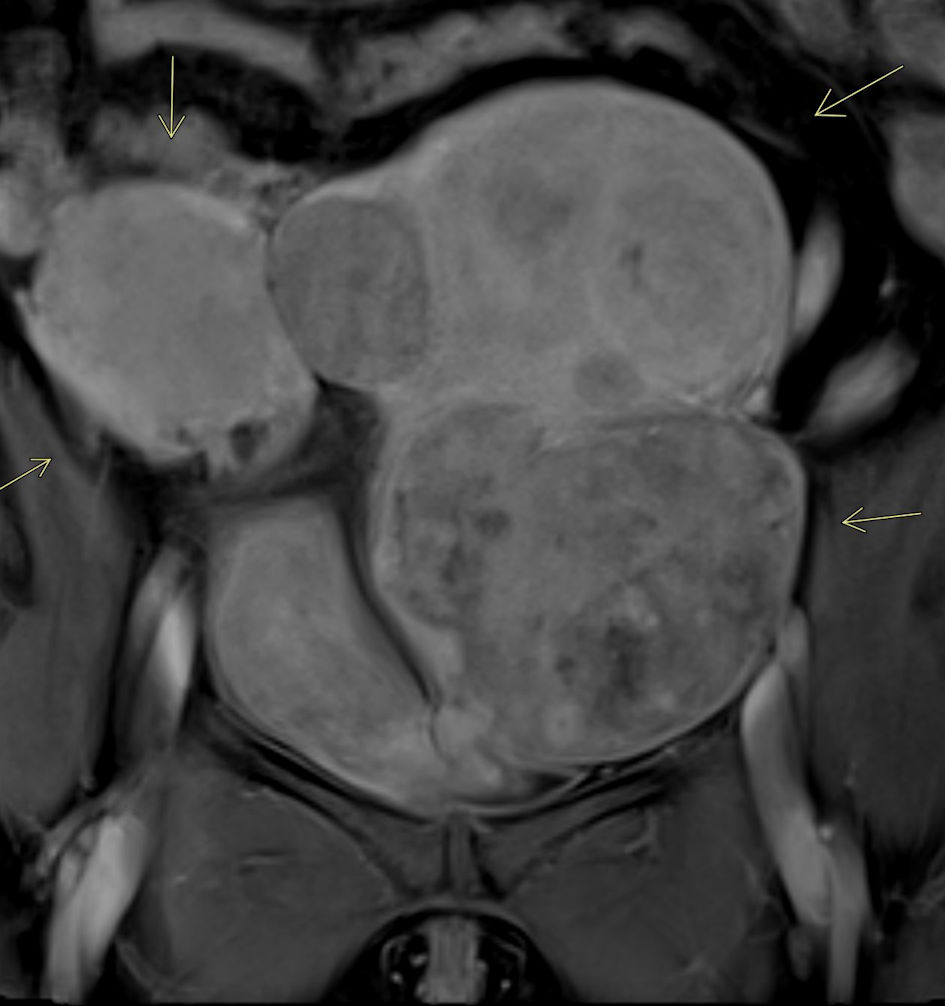
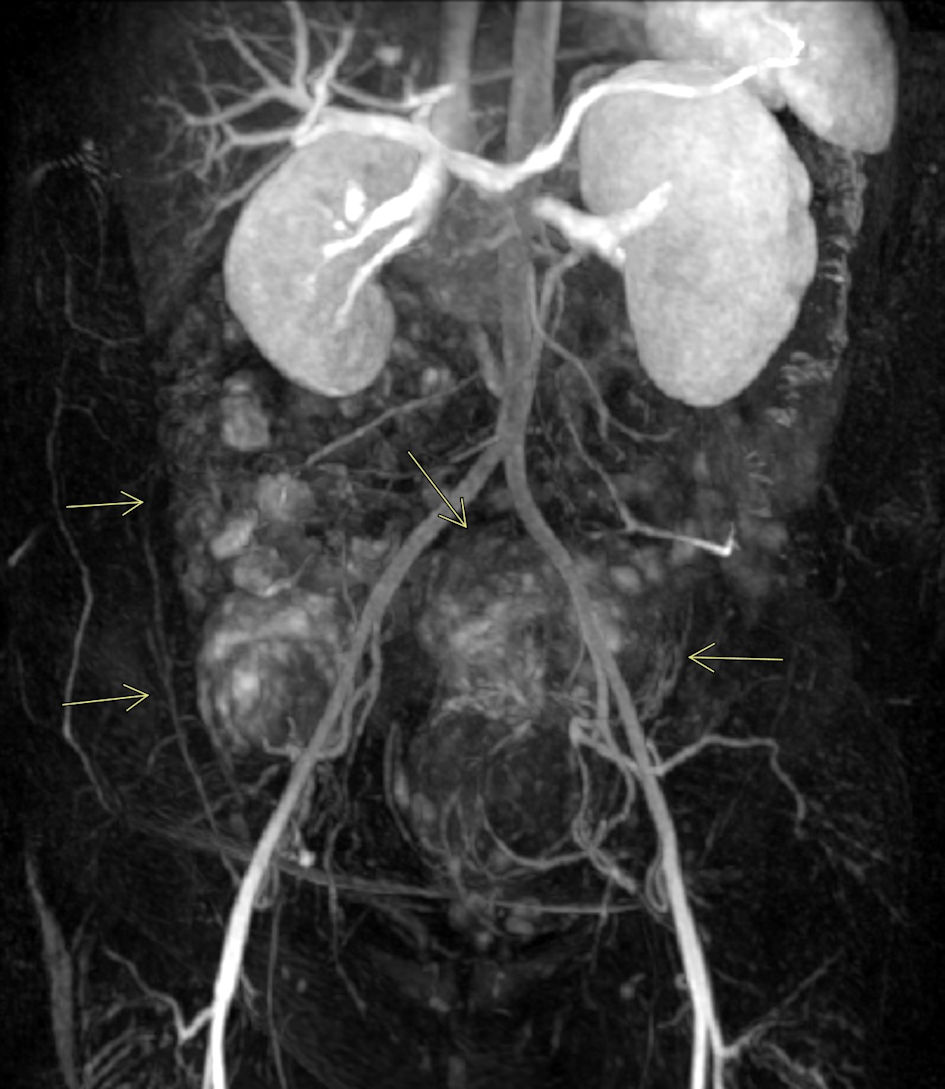
A magnetic resonance imaging (MRI) was subsequently ordered to further evaluate the extent of her uterine fibroids and anatomical variation due to uterine didelphys. MRI confirmed the presence of uterine didelphys with multiple large enhancing uterine fibroids distorting the endometrial cavity (Fig. 1). Fibroids were identified in both uteri, with the largest on the left measuring 14 × 11 × 11 cm and the largest on the right measuring 5.8 × 5.5 × 4.7 cm. Magnetic resonance angiography (MRA) demonstrated normal pelvic arterial anatomy with bilaterally enlarged uterine arteries, more prominent on the left. Prior to uterine artery fibroid embolization, MRA provides excellent visualization of the pelvic anatomy, assisting the interventionist in pre-procedural planning for the procedure (Fig. 2).
 Click for large image |
Figure 1. MRI of pelvis with intravenous contrast. Contrast-enhanced MRI of the pelvis demonstrates uterine didelphys with multiple large uterine fibroids, resulting in significant distortion of the endometrial cavity (arrows). |
 Click for large image |
Figure 2. MRA of pelvis. Pelvic MRA confirms the highly vascular nature of the uterine fibroids, particularly notable in this case involving multiple large fibroids affecting both uteri (arrows). |
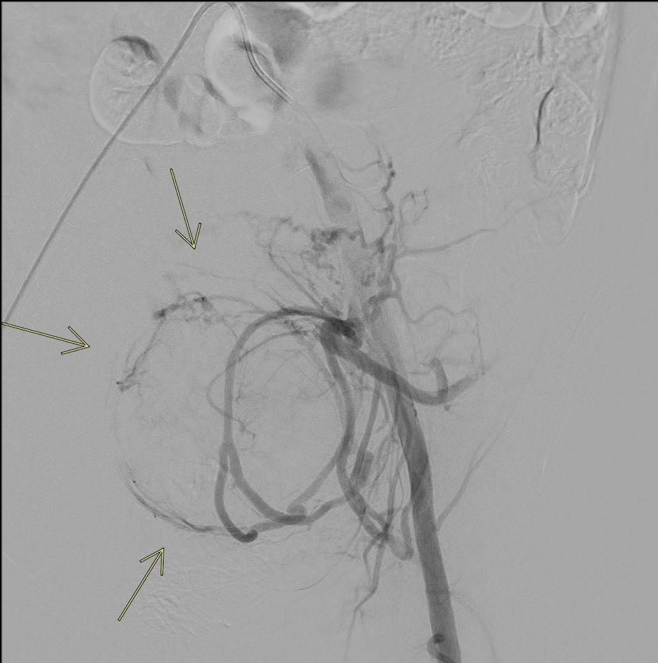
UFE was then performed using standard interventional radiology technique. Moderate sedation was given using Versed (Pfizer Inc. New York, NY) 1 mg intravenous (IV) and fentanyl (Janssen Pharmaceutical Athens, GA) 50 µg IV. A conventional aortic angiogram and a selective iliac artery angiogram were performed. The left uterine arteriogram demonstrated a prominent vascular blush consistent with large fibroids, correlating with prior MRI findings. A single, enlarged left uterine artery was noted supplying the markedly enlarged fibroid uterus. A TriSalus TriNav Infusion System (Trisalus Life Science Westminster, CO) antireflux infusion microcatheter was placed through the diagnostic catheter and advanced into the left uterine artery, and selective arteriogram was performed (Fig. 3). Intra-arterial embolization was performed with Terumo Hydropearl 200 - 400 µm microspheres.
 Click for large image |
Figure 3. Selective left iliac conventional angiogram. Left iliac artery angiogram demonstrates prominent hypervascular fibroids occupying a substantial portion of the left uterus (arrows). |
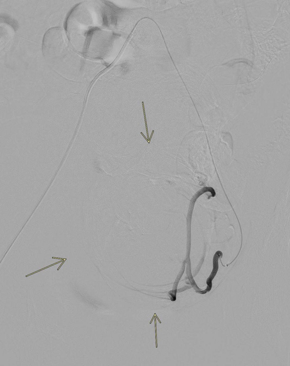
Post-embolization imaging of the left internal iliac artery showed no opacification of intrauterine branches, indicating a successful angiographic result (Fig. 4). The right uterine arteriogram also demonstrated standard anatomy, along with a significant vascular blush consistent with large fibroids. The diagnostic catheter was retracted into the right internal iliac artery, and selective arteriogram was performed. The microcatheter was advanced into the right uterine artery, and intra-arterial embolization was performed by injecting Terumo Hydropearl 200 - 400 µm microspheres. Post-embolization imaging of the right uterine artery showed no opacification of intrauterine branches, confirming successful embolization.
 Click for large image |
Figure 4. Post-embolization completion angiogram. Left uterine artery angiogram following embolization demonstrates no residual flow to the large fibroids, correlating with excellent radiologic and clinical outcomes (arrows). |
Four weeks after undergoing UFE, the patient was contacted by phone for a follow-up interview. She reported doing very well, with a significant reduction in abnormal bleeding as well as a marked decrease in pressure and pain caused by the fibroids. The symptoms that initially prompted her to consult her gynecologist have now resolved. A pelvic MRI is scheduled 3 months post-procedure to further evaluate her progress.
| Discussion | ▴Top |
Uterine anomalies are present in approximately 3-5% of women. Other common congenital anomalies include septate uterus, bicornuate uterus, and unicornuate uterus. A septate uterus has a normal external contour but contains a fibrous or muscular septum dividing the endometrial cavity. A unicornuate uterus results from incomplete development of one Mullerian duct, leading to a single uterine horn. A bicornuate uterus presents with an abnormal external shape and two endometrial cavities due to partial failure of fusion of the Mullerian ducts [6]. While many uterine anomalies are asymptomatic, some can be associated with recurrent miscarriage, pelvic pain, or abnormal menstrual bleeding. These anomalies are often not identified until pregnancy, when they may be discovered incidentally on imaging such as ultrasound.
The exact cause of uterine fibroids remains unclear; however, several factors are believed to contribute to their development. Genetic mutations, elevated levels of estrogen, progesterone, or certain growth hormones have all been implicated in fibroid formation. Among various risk factors, vitamin D deficiency has emerged as one of the most significant. Low vitamin D levels are associated with larger fibroid size, likely due to the vitamin’s role in regulating cell growth and differentiation across multiple tissues, including those of the reproductive system. Ethnicity also appears to influence fibroid risk [7]. African American women have both the highest incidence of uterine fibroids and the lowest average vitamin D levels. Additionally, Vietnamese women - part of the Southeast Asian population - are 29% more likely to be diagnosed with fibroids compared to Caucasian women. This group ranks fourth in incidence among all ethnicities [8]. These findings help inform clinicians of demographic trends and potential risk factors to consider during diagnosis and treatment planning.
Annually, there are significantly more hysterectomies than UFEs performed. There are approximately 600,000 hysterectomies performed, and about 14,000 UFE procedures in the United States [9]. Uterine-sparing treatments for uterine fibroids include myomectomy, which preserves fertility by removing only the leiomyomas and noninvasive treatments endometrial ablation and hysterectomy, which involves the complete removal of the uterus. Other treatment options for uterine fibroids include prescription medications, noninvasive procedures, and additional minimally invasive techniques. Medical therapies such as gonadotropin-releasing hormone (GnRH) agonists, GnRH antagonists, and intrauterine devices (IUDs) can help manage fibroid-related symptoms and reduce fibroid size, though they do not eliminate the fibroids entirely. MRI-guided focused ultrasound surgery (MRgFUS) is a noninvasive treatment that uses MRI to precisely locate fibroids and high-frequency sound waves to thermally ablate fibroid tissue. This procedure is relatively safe and has shown good outcomes in symptom relief. Another minimally invasive alternative to UFE is radiofrequency ablation, which uses targeted heat to destroy fibroid tissue and reduce blood supply to the fibroids, leading to shrinkage [10]. All treatment options are discussed with the patient, and the most appropriate plan is selected based on clinical findings and patient preference.
UFE is an effective technique that targets the small blood vessels supplying uterine fibroids. Embolic beads are selected based on vessel size: smaller beads penetrate deeper into the fibroid to induce central necrosis, while larger beads occlude vessels more proximally. The beads typically range from 75 to 600 µm. In this case, a combination of 200 - 400 µm beads were used, which is standard practice for UFE [11]. These are “bland” embolic microspheres, meaning they do not have chemotherapeutic agents attached to them when used in locoregional treatments for malignant liver tumors [12]. Since uterine fibroids are benign, bland embolization is both appropriate and preferred, avoiding the systemic side effects associated with drug-eluting beads. As with any arterial embolization procedure, an expected post-procedural treatment response known as post-embolization syndrome (PES) can occur, which is a transient condition characterized by low-grade fever, localized pelvic pain, and malaise, typically without leukocytosis. This self-limiting syndrome generally resolves within 24 h and is managed with anti-inflammatory medications such as Toradol (Roche Laboratories Branchburg, NJ) and, when needed, narcotics like Demerol (Pfizer Inc. New York, NY) for pain control. Overall, most patients can be discharged from the post-procedural unit the same day or after an overnight observation, depending on symptom severity. Follow-up imaging with pelvic MRI is typically performed at 3 months and 1 year post-procedure to monitor fibroid shrinkage. In rare cases, intracavitary submucosal fibroids may undergo expulsion following embolization. However, this risk is significantly reduced by performing pre-procedural MRI, which helps identify and exclude patients with intracavitary fibroids from undergoing UFE.
In general, fibroid recurrence after UFE is well documented, and the rate is higher compared to hysterectomy and myomectomy. Recurrence rates vary depending on the embolization technique used and the initial size of the fibroids. Fibroid size and number are factors for recurrence, but the overall symptom recurrence rate is about 17% at 30 months post-embolization [9]. Failure of incomplete embolization is possible, presenting with growth of fibroids or residual fibroids. These patients present with pain from sloughing fibroid material. Despite the risk of recurrence, UFE has been shown to improve uterine blood flow, particularly in patients with adenomyosis, leading to symptomatic relief when fibroids coexist with this condition. After UFE, healthy uterine tissue remains unaffected, demonstrating the efficacy of this treatment.
Fertility outcomes following UFE continue to be extensively studied. Published data suggest that approximately 38.3% of women attempting pregnancy after UFE achieve successful conception, although this figure varies across studies due to differing patient populations and follow-up durations. In comparison, myomectomy demonstrates higher reported fertility rates, ranging between 53% and 56%, making it a preferred option for women with fertility concerns [10]. Despite UFE’s advantages in symptom management, with reported symptom improvement rates exceeding 85% and significantly shorter hospital stays averaging 1 - 2 days compared to 3 - 5 days for myomectomy, pregnancy outcomes following UFE are less favorable. Studies indicate that UFE is associated with increased risks of preterm delivery (up to 20% in some cohorts versus approximately 10-12% post-myomectomy) and spontaneous abortion rates as high as 25%, compared to 15-20% after myomectomy [11]. Additionally, incidences of low birth weight and placental abnormalities have been reported more frequently post-UFE. These findings emphasize the need for comprehensive counseling for women of reproductive age considering UFE, balancing the benefits of minimally invasive treatment and symptom relief against potential risks to future pregnancy outcomes. Larger prospective studies are needed to better quantify fertility rates and optimize patient selection for UFE.
Since 2016, the overall number of UFE procedures has steadily increased, reflecting growing recognition of UFE as a viable nonsurgical treatment option for uterine fibroids. However, significant disparities remain in the availability and utilization of UFE across different communities. Urban hospitals are substantially more likely to perform UFE compared to surgical interventions such as hysterectomy, with urban centers reporting UFE rates up to 30-40% higher than rural facilities. In contrast, rural and small hospitals particularly those serving populations between 10,000 and 50,000 continue to underutilize UFE, often defaulting to surgical approaches [12]. This disparity is attributed to factors such as limited access to interventional radiology specialists, resource constraints, and differences in patient awareness. Regional variations are also notable: hysterectomy rates are highest in the East South-Central region, with rates exceeding the national average by approximately 15%, while UFE procedures remain relatively low nationwide. The majority of UFE treatments are concentrated in the West North Central and Mountain regions, where interventional radiology services are more accessible and integrated into patient care pathways. These patterns underscore a critical need for targeted efforts to expand UFE availability and education in underserved rural areas and high-hysterectomy regions to promote more equitable, less invasive treatment options.
Acknowledgments
None to declare.
Financial Disclosure
None to declare.
Conflict of Interest
None to declare.
Informed Consent
Informed consent was obtained.
Author Contributions
Nicole T. Stambo, the corresponding author, wrote and researched the entity for this case report as well as being present in the interventional radiology (IR) suite during the procedure. Dr. Glenn W. Stambo provided the patient and performed the procedure.
Data Availability
The data supporting the findings of this study are available from the corresponding author upon reasonable request.
| References | ▴Top |
- Rezai S, Bisram P, Lora Alcantara I, Upadhyay R, Lara C, Elmadjian M.
Didelphys uterus: a case report and review of the literature. Case Rep Obstet Gynecol.
2015;2015:865821.
doi pubmed - Defran AJ, Forestier C, Morgan E, Thomas M. Uterine leiomyoma in the
context of uterine didelphys: a case report. Cureus. 2023;15(9):e44791.
doi pubmed - Mashala JN, Wekesa D, Chemwey R, Pulei A, Kihara A. An incidental case of uterus didelphys and fibroids: Right sided myomectomy and left hemi hysterectomy. Anatomy Journal of Africa. 2020;9(2):1762-1764.
- Kroncke T. An update on uterine artery embolization for uterine
leiomyomata and adenomyosis of the uterus. Br J Radiol.
2023;96(1143):20220121.
doi pubmed - Kim MD. Uterine artery embolization for leiomyomas and adenomyosis: A
pictorial essay based on our experience from 1300 cases. Korean J Radiol.
2019;10:1462-1473.
doi pubmed - Kaufman CD. Currently available embolics for uterine fibroid embolization. Endovascular Today. 2020. https://evtoday.com/articles/2020-apr/currently-available-embolics-for-uterine-fibroid-embolization.
- Stambo G, Cragan D. Response rates of hepatocellular carcinoma and hepatic colorectal cancer metastases to drug eluting bead regional liver therapy. Hepatoma Research. https://www.oaepublish.com/articles/2394-5079.2017.12.
- Svarc P, Taudorf M, Nielsen MB, Stroomberg HV, Roder MA, Lonn L.
Postembolization syndrome after prostatic artery embolization: a systematic review. Diagnostics
(Basel). 2020;10(9):659.
doi pubmed - Marret H, Cottier JP, Alonso AM, Giraudeau B, Body G, Herbreteau D.
Predictive factors for fibroids recurrence after uterine artery embolisation. BJOG.
2005;112(4):461-465.
doi pubmed - Saha S, Feldman M, Ullman R, Korach K. A systematic review of the
effect of women’s choice of health facility on delivery outcomes. BMC Pregnancy and
Childbirth. 2019;19(1):319.
doi - Vedantham S, Sterling KM, Goodwin SC, Spies JB, Shlansky-Goldberg R,
Worthington-Kirsch RL, Andrews RT, et al. I. Uterine fibroid embolization: preprocedure
assessment. Tech Vasc Interv Radiol. 2002;5(1):2-16.
doi pubmed - Elhakim TS, Smolinski-Zhao S, Miyasato D, Lee V, Mansur A, Puello M,
Mercaldo N, et al. Disparities in utilization of uterine fibroid embolization. JAMA Netw Open.
2025;8(9):e2532100.
doi pubmed
This
article is distributed under the terms of the Creative Commons Attribution Non-Commercial 4.0
International License, which permits unrestricted non-commercial use, distribution, and
reproduction in any medium, provided the original work is properly cited.
Journal
of Clinical Gynecology and Obstetrics is published by Elmer Press Inc.