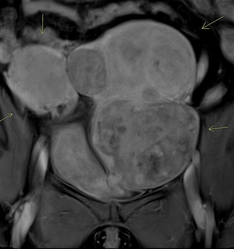
Journal of Clinical Gynecology and Obstetrics, ISSN 1927-1271 print, 1927-128X online, Open Access
Article copyright, the authors; Journal compilation copyright, J Clin Gynecol Obstet and Elmer Press Inc
Journal website https://jcgo.elmerpub.com

Case Report

Volume 14, Number 4, December 2025, pages 184-188


Uterine Fibroid Embolization in Patient With Symptomatic Uterine Leiomyomas in Uterine Didelphys

Figures

Figure 1.
Figure 1. MRI of pelvis with intravenous contrast. Contrast-enhanced MRI of the pelvis demonstrates uterine didelphys with multiple large uterine fibroids, resulting in significant distortion of the endometrial cavity (arrows).
Figure 2.
Figure 2. MRA of pelvis. Pelvic MRA confirms the highly vascular nature of the uterine fibroids, particularly notable in this case involving multiple large fibroids affecting both uteri (arrows).
Figure 3.
Figure 3. Selective left iliac conventional angiogram. Left iliac artery angiogram demonstrates prominent hypervascular fibroids occupying a substantial portion of the left uterus (arrows).
Figure 4.
Figure 4. Post-embolization completion angiogram. Left uterine artery angiogram following embolization demonstrates no residual flow to the large fibroids, correlating with excellent radiologic and clinical outcomes (arrows).