| Journal of Clinical Gynecology and Obstetrics, ISSN 1927-1271 print, 1927-128X online, Open Access |
| Article copyright, the authors; Journal compilation copyright, J Clin Gynecol Obstet and Elmer Press Inc |
| Journal website https://jcgo.elmerpub.com |
Original Article
Volume 000, Number 000, March 2025, pages 000-000
Magnetic Resonance Imaging-Based Prediction of Uterine Fibroid Size Reduction by Relugolix Using T2-Weighted Signal Intensity Ratio: A Retrospective Multicenter Study
Jun Matsukawaa, d , Toshitada Hirakab, Akiko Sugiyamac, Hiromu Kanekoa, Fumihiro Nakamuraa, Nanako Nakaia, Kyoko Takahashia, Isao Takeharaa, Masafumi Kanotob, Satoru Nagasea
aDepartment of Obstetrics and Gynecology, Yamagata University Faculty of Medicine, Yamagata 990-9585, Japan
bDepartment of Radiology, Division of Diagnostic Radiology, Yamagata University Faculty of Medicine, Yamagata 990-9585, Japan
cDepartment of Obstetrics and Gynecology, Yamagata Saisei Hospital, Yamagata 990-0818, Japan
dCorresponding Author: Jun Matsukawa, Department of Obstetrics and Gynecology, Yamagata University Faculty of Medicine, Yamagata 990-9585, Japan
Manuscript submitted January 13, 2025, accepted March 17, 2025, published online March 25, 2025
Short title: Prediction of Uterine Fibroid Reduction by Relugolix
doi: https://doi.org/10.14740/jcgo1506
| Abstract | ▴Top |
Background: The aim of this study was to determine whether T2-weighted magnetic resonance imaging (MRI) can predict the volume reduction effect of relugolix on uterine fibroids by evaluating the signal intensity ratio between uterine fibroids and the iliopsoas muscle.
Methods: This multicenter retrospective study included patients who underwent surgery for uterine fibroids at Yamagata University Hospital and Yamagata Saisei Hospital between January 2020 and December 2021. Patients received daily oral relugolix (40 mg) from the first day of menstruation after their first consultation until the day before surgery. MRI was performed before treatment and 1 - 2 weeks before surgery to assess fibroid size and signal intensity on T2-weighted images (T2WIs). The signal intensity ratio (T2WI ratio) of fibroids to iliac muscle was used to ensure consistency. Patients were classified based on a 50% fibroid volume reduction, and T2WI ratios were compared. Furthermore, receiver operating characteristic (ROC) analysis was conducted to determine the optimal T2WI ratio cutoff for predicting volume reduction.
Results: Overall, 39 patients with 70 uterine fibroids were included in the statistical analysis. There was no significant difference in the duration of relugolix treatment between the groups. The group with ≥ 50% volume reduction had significantly larger T2WI ratios than the group with < 50% volume reduction (P = 0.006). In the ROC curve analysis, a T2WI ratio cutoff value of 1.751 was determined to be positive when considering a volume reduction rate of ≥ 50%. The corresponding area under the curve was 0.772, indicating moderate accuracy. The negative predictive value was high in this case, reaching 0.974.
Conclusion: T2WI ratio may be useful for predicting the volume-reducing effect of relugolix on uterine fibroids.
Keywords: MRI; Relugolix; Uterine fibroids; Uterine leiomyoma; Volume reduction
| Introduction | ▴Top |
Uterine fibroids are the most common benign tumors of the uterus and are clinically apparent in about 25% of women [1]. Careful pathological examination of surgical specimens suggests that the prevalence is as high as 77% [2]. They present with various symptoms depending on their location, size, and number. Uterine fibroids can reduce a woman’s quality of life by causing excessive menstruation and dysmenorrhea [1], and can lead to complications such as infertility, miscarriage, and premature delivery [3, 4], making them a substantial concern, especially in reproductive and perinatal medicine.
In 2019, the world’s first oral gonadotropin-releasing hormone (GnRH) antagonist, relugolix, was launched in Japan, and has recently been increasingly used as a hormone therapy for uterine fibroids. Unlike conventional GnRH agonist drugs, relugolix does not cause a transient increase in estrogen; rather, it causes a steep decrease in estrogen, and is expected to have a faster effect on the various symptoms caused by uterine fibroids [5]. However, side effects such as hot flashes and uterine bleeding are frequently observed [6], and, as with GnRH agonists, long-term administration may cause serious complications, such as osteoporosis [7]. In clinical practice, relugolix is increasingly being administered to patients seeking minimally invasive hysterectomy or myomectomy in the hope of reducing the size of the entire uterus or uterine fibroids. However, even with the administration of relugolix, some uterine fibroids fail to shrink. Administration of the drug to such patients wastes their precious time and money.
Several studies have been reported to predict the effect of GnRH agonists on shrinking uterine fibroids, many of which use magnetic resonance imaging (MRI). By classifying T2-weighted MRIs of uterine fibroids and correlating them with pathological findings, it was reported that homogeneous uterine fibroids with high T2 signal intensity have high cell density and proliferation ability, and shrink significantly with GnRH agonists [8, 9]. Recent studies have reported that fibroids with mediator complex subunit 12 (MED12) gene mutations, which are found in approximately 70% of uterine fibroids [10], show low T2 signal intensity and limited shrinkage with GnRH agonists [11]. However, no studies have been reported yet on the predictive effect of relugolix on reducing uterine fibroid size. The aim of this study was to investigate the feasibility of predicting the effect of relugolix on uterine fibroid volume reduction through the analysis of pretreatment MRI scans. By establishing a predictive model, we can potentially avoid unnecessary administration of relugolix in cases where the anticipated effect is low, leading to more efficient and personalized treatment approaches.
| Materials and Methods | ▴Top |
This study was approved by the Ethics Committee of the Yamagata University Faculty of Medicine (approval number 2021-102). This study was conducted in compliance with the ethical standards of the responsible institution on human subjects as well as with the Helsinki Declaration. This study was a multicenter, retrospective study; it included patients who underwent surgery for uterine fibroids at Yamagata University Hospital and Yamagata Saisei Hospital between January 2020 and December 2021. The patient was administered relugolix 40 mg/day orally every day from the first day of menstruation after the first consultation until the day before surgery. Patients underwent MRI before starting relugolix treatment and 1 to 2 weeks before surgery to assess fibroid size and signal intensity on T2-weighted images (T2WIs).
Each patient’s evaluation focused on up to three uterine fibroids with a major diameter of ≥ 3 cm prior to relugolix administration. The exclusion criteria were: patients who did not have uterine fibroids with a major diameter of ≥ 3 cm, those who received relugolix for < 30 days, and those with a pretreatment MRI date ≥ 90 days before the start of relugolix administration.
All MRI scans were acquired using standard well-established imaging parameters and protocols. The slice thicknesses for the axial and sagittal images were 4 - 13 and 4 - 12 mm, respectively. Fibroid volume and signal intensity on T2WIs were determined using Picture Archiving and Communication System (PACS) (Ev Insite, PSP Corp., Tokyo, Japan).
To ensure consistency and comparability, the signal intensity on T2WIs was measured as a relative ratio value rather than an absolute value. In general, in radiology research, the absolute value of the signal intensity of T2WIs varies depending on the imaging parameters and imaging device, making them unsuitable for image analysis using image data from multiple facilities, as in this study. Specifically, using the T2-weighted MRI taken before treatment, the ratio of the signal intensity of the targeted fibroid to that of the iliac muscle was calculated as the “T2WI ratio.” The signal intensity ratio of uterine fibroids and iliac muscles on T2WIs can be easily measured, and is a method that can be used in routine clinical practice. There are several reports that the signal intensity ratio of uterine fibroids and iliac muscles on T2WIs is useful for predicting the effect of fibroid volume reduction in uterine artery embolization [12, 13]. The uterine fibroid signal intensity was determined using the region of interest (ROI), which was set as wide as possible to avoid obvious cystic or necrotic regions, and the average signal intensity within the ROI was adopted. The signal intensity of the iliac muscle was determined similarly (Fig. 1). The volume reduction rate of the uterine fibroids was determined using MRI scans before and after treatment with relugolix. Specifically, it was calculated as the ratio between the products of the three vertical diameters of the fibroids before and after treatment. In these image analyses, the T2WI ratio was measured by a board-certified radiologist with more than 10 years of experience in obstetrics and gynecological imaging, and the volume reduction rate of uterine fibroids was measured by a gynecologist. These specialists performed the measurements without knowing each other’s image analysis results.
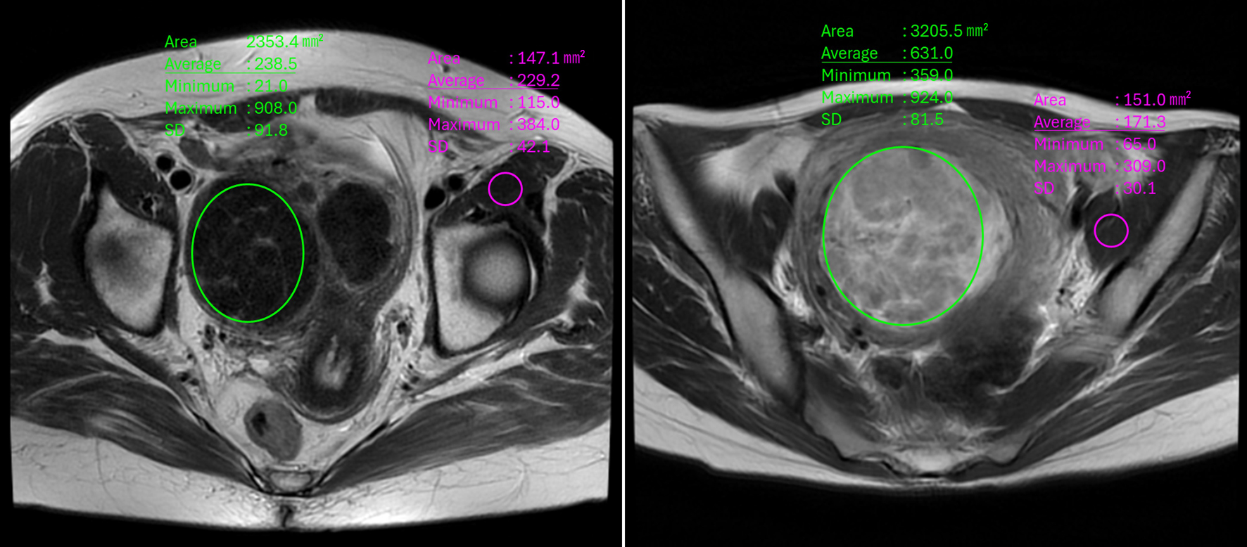 Click for large image | Figure 1. Region of interest (ROI) setting in T2-weighted images. In the T2-weighted image of this case, the ROI of the uterine fibroid was set in the area surrounded by the green line. We selected a site that contained as large an ellipse as possible without including cystic or necrotic areas with high signal intensity. The ROI was set for the iliac muscle in the same way (the area surrounded by the purple line). |
First, the uterine fibroids of the eligible patients were divided into two groups based on the 50% reduction rate of uterine fibroid volume, and the T2WI ratios between the groups were compared (Mann-Whitney U test). Second, receiver operating characteristic (ROC) curves were constructed to determine the cutoff value of T2WI ratio for fibroid volume reduction. In addition, correlations between the two continuous variables, volume loss rate and T2WI ratio, were evaluated using Spearman’s rank correlation coefficient. Statistical analysis was performed using Easy R ver. 1.54 (Jichi Medical University, Ibaraki, Japan).
| Results | ▴Top |
Of the 49 eligible patients, 10 were excluded based on the exclusion criteria. Therefore, a total of 39 patients with 70 uterine fibroids were included for statistical analysis (Fig. 2). The median age of the eligible patients was 43 (interquartile range: 37.5 - 46) years, and the median duration of relugolix treatment was 97 (interquartile range: 80 - 110) days. Table 1 shows the characteristics of each group divided by a uterine fibroid volume reduction rate of 50%. There were no significant differences in age, body mass index, pregnancy, or duration of relugolix treatment (t-test).
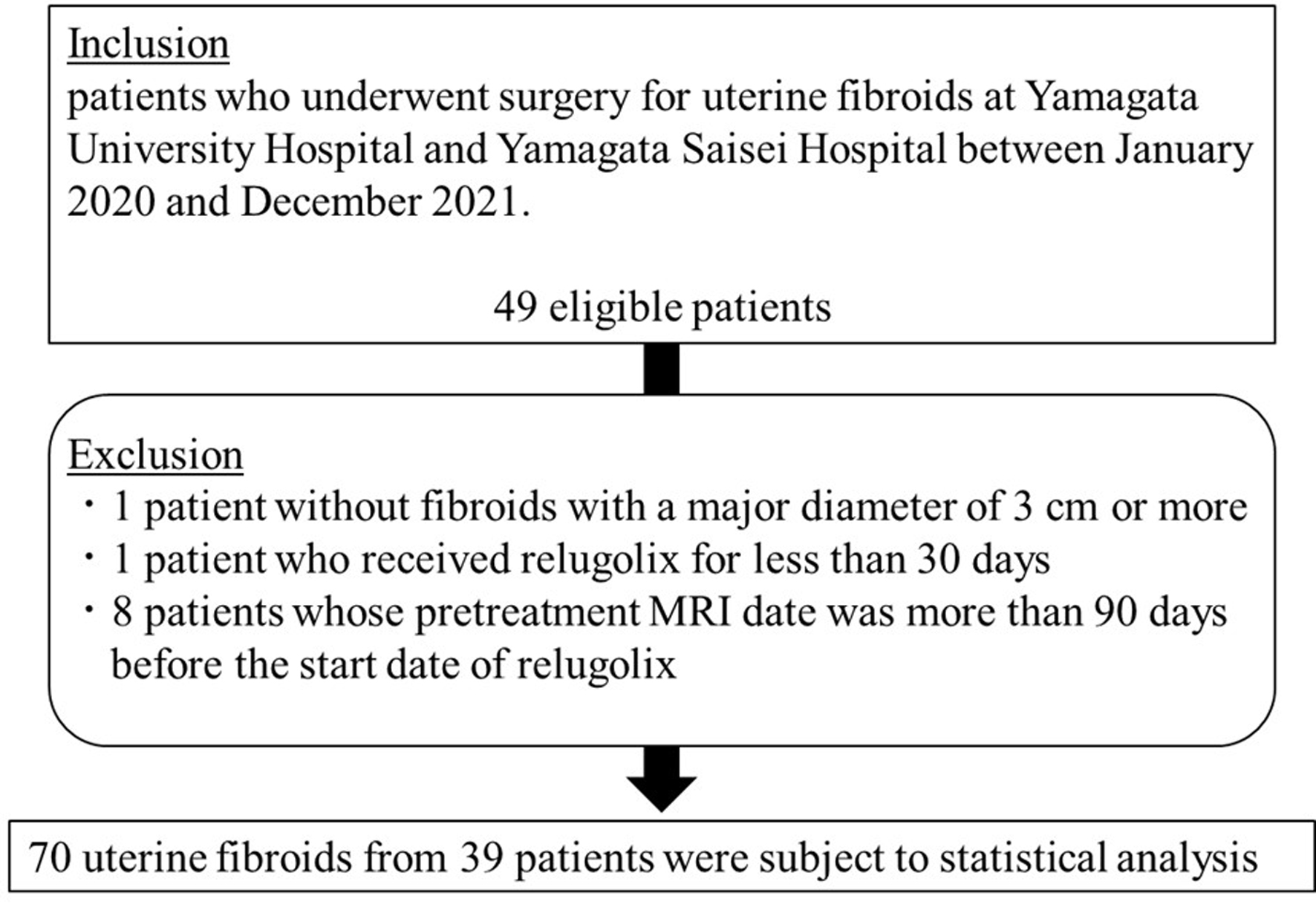 Click for large image | Figure 2. Patients and uterine fibroids selection criteria. |
 Click to view | Table 1. Characteristics of Uterine Fibroids Divided by Volume Reduction Rate of 50% |
Uterine fibroids that met the criteria were divided into two groups based on a 50% volume reduction rate, and box plots were created for the T2WI ratios of uterine fibroids in each group to compare them between the groups. The T2WI ratio was significantly larger in the group with a volume reduction rate of ≥ 50% than in the group with a volume reduction rate of < 50% (P = 0.006) (Fig. 3).
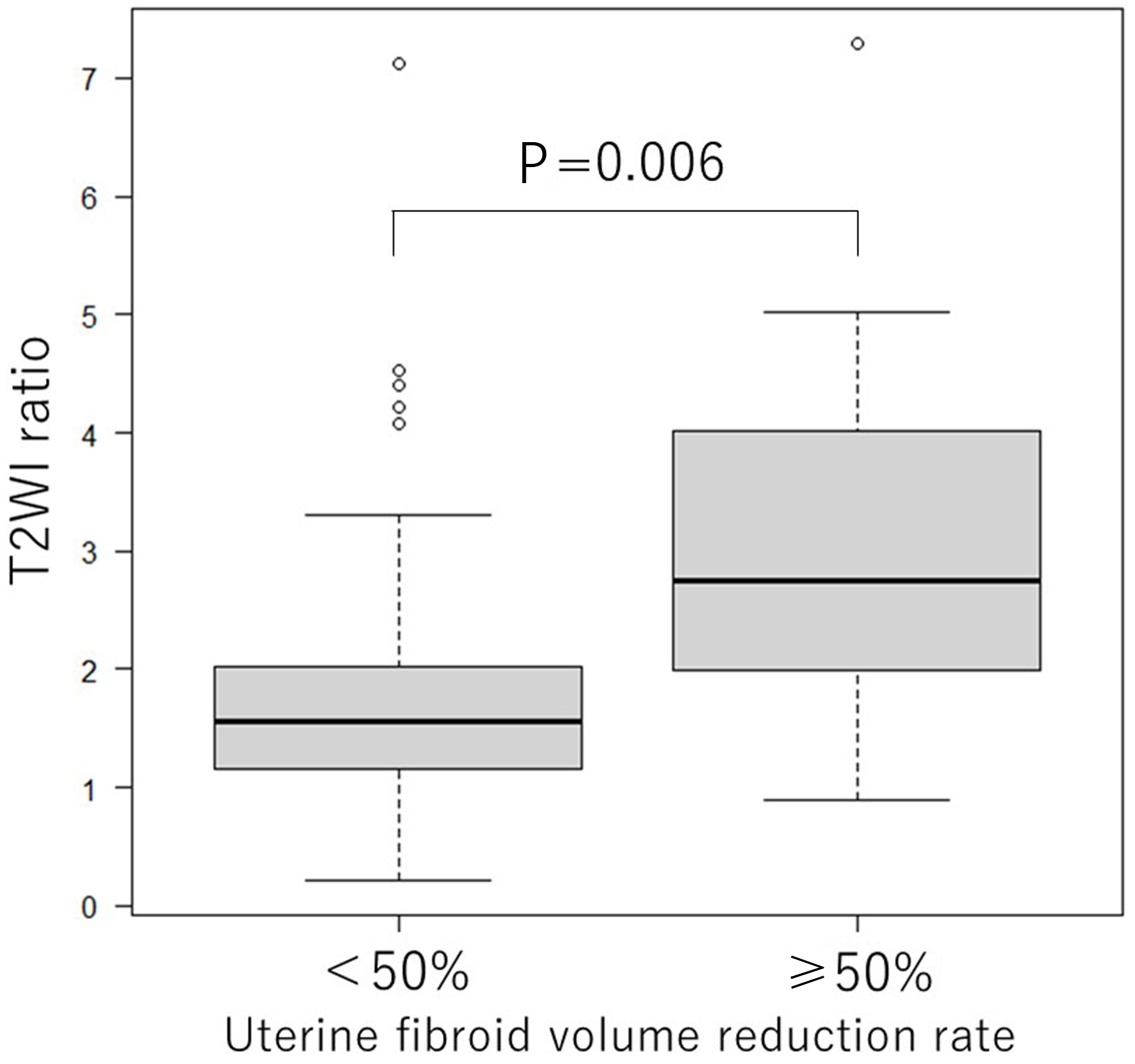 Click for large image | Figure 3. Boxplot comparing T2WI ratio by dividing uterine fibroid volume reduction rate by 50%. T2WI ratio is significantly larger in the group with a volume reduction of ≥ 50% compared to the group with a volume reduction of < 50% (P = 0.006). T2WI: T2-weighted image. |
In addition, ROC curve was constructed to determine the cutoff value of the T2WI ratio, with a volume reduction rate of ≥ 50% assumed to be effective (positive) (Fig. 4). The cutoff value was 1.751 and the area under the curve was 0.772 (95% confidence interval: 0.598 - 0.945). The accuracy of the test was also analyzed when the T2WI ratio cutoff value was set at 1.751 (Table 2). Notably, the negative predictive value was 0.974, which meant that if the T2WI ratio was less than 1.751, there was a 97.4% probability of a < 50% reduction in fibroid volume. The positive predictive value was 0.290.
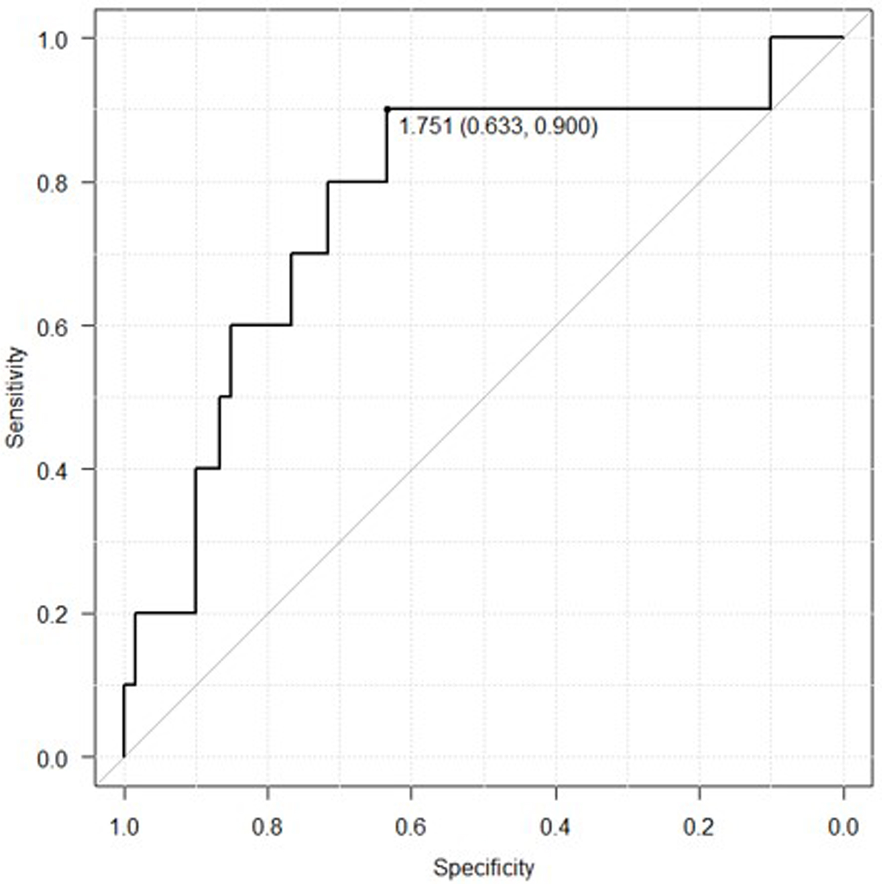 Click for large image | Figure 4. Receiver operating characteristic curve created by assuming that a fibroid volume reduction rate of 50% or more is effective (positive). The cut-off value was 1.751 and the area under the curve was 0.772 (95% CI, 0.598 - 0.945). CI: confidence interval. |
 Click to view | Table 2. Evaluation of Test Accuracy When T2-Weighted Image Ratio Cutoff Value Is Set to 1.751 |
Furthermore, correlation between the two continuous variables, volume loss rate and T2WI ratio, was evaluated (Fig. 5). The Spearman’s rank correlation coefficient was 0.222, and although there was a slightly positive correlation between the volume reduction rate and the T2WI ratio, it was not significant (P = 0.0647).
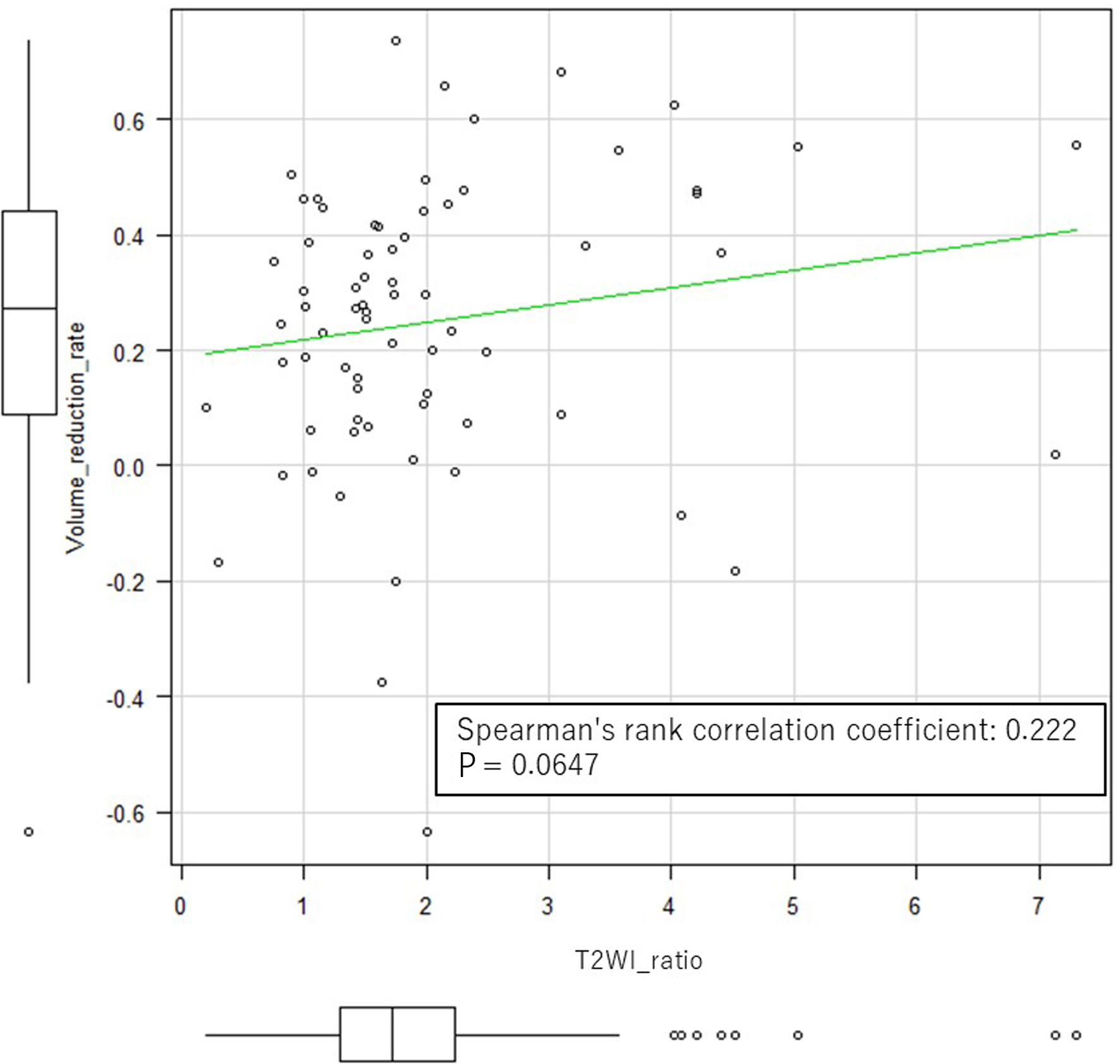 Click for large image | Figure 5. Spearman’s rank correlation coefficient determined for two continuous variables: the T2WI ratio and the rate of uterine fibroid volume reduction. T2WI: T2-weighted image. |
| Discussion | ▴Top |
This study suggested that the T2WI ratio may be used to predict the volume reduction effect of relugolix on uterine fibroids. Relugolix has been reported to be effective in reducing uterine fibroid volume; however, uterine fibroids that do not show this effect are often encountered. If the effect of volume reduction can be predicted before treatment, relugolix can be aggressively administered to more suitable cases, and other drug treatments or early surgery can be recommended to unsuitable ones. Since a significant difference was observed in the T2WI ratio between the groups divided by 50% volume reduction rate, the T2WI ratio may be a useful criterion for determining whether or not to use relugolix for the purpose of reducing uterine fibroids. In addition, a cutoff value of 1.751 for the T2WI ratio was derived from the ROC curve, which was created assuming that a volume reduction rate of 50% or more is effective when relugolix is administered. The area under the ROC curve was 0.772 (95% confidence interval: 0.598 - 0.945), indicating moderate accuracy. When the cutoff value of the T2WI ratio was set at 1.751, the negative predictive value was particularly high at 0.974, so this cutoff value may be useful for selecting uterine fibroids for which relugolix has a low volume reduction effect. However, it should be noted that this cutoff value is not predictive of higher uterine fibroid volume reduction efficacy, as the positive predictive value was low at 0.290.
Regarding GnRH agonists, there are several reports discussing the prediction of the uterine fibroid volume reduction effect, and many of them have used MRI. Matsuno et al classified T2-weighted MRIs of uterine fibroids into three types, matched them with pathological findings, and verified whether the effect of GnRH agonist on reducing fibroid volume could be predicted. They reported that homogeneous hyperintense uterine fibroids on T2WIs were histopathologically identified as cellular leiomyoma, and that administration of a GnRH agonist resulted in a greater reduction in fibroid volume in such cases [8]. In addition, Oguchi et al reported that cell density and proliferative activity in fibroid tissue tended to increase in response to increased signal intensity on T2WIs of uterine fibroids. They suggested that these factors were involved in the uterine fibroid volume reduction effect of GnRH agonists [9]. Recent reports indicate that uterine fibroids with mutations in the MED12 gene, which is found in 70% of uterine fibroids [10], exhibit limited volume reduction when treated with GnRH agonists and low signal intensity on T2WIs [11]. Contrary to previous studies, ours used a continuous variable: the intensity ratio of fibroids to iliac muscles. In radiology research, the absolute signal intensity of T2WIs varies with imaging parameters and devices, making cross-facility image analysis challenging. Therefore, MRI signal intensity is generally used as a relative ratio value rather than an absolute value. This study also adopted this method to maintain consistency with established practices in the field. Measuring the signal intensity of uterine fibroids and iliac muscles on T2WIs using ROI and calculating the ratio is a simple and common clinical practice [12, 13].
In this study, we divided the rate of uterine fibroid volume reduction by 50% for analysis, but there is no consensus on the extent to which the uterine fibroid volume should be reduced to obtain a satisfactory clinical outcome. Spies et al reported that patients with a < 30.5% uterine fibroid volume loss were more than three times more likely to be dissatisfied than those with a > 56.3% volume loss [14]. Hald et al also reported significant symptom recurrence in patients with < 24% uterine fibroid volume loss compared to those with > 48% uterine fibroid volume loss [15]. Moreover, Duvnjak et al stated that a > 50% reduction in the uterine fibroid volume is an appropriate outcome [16]. Based on these reports, it can be said that the value of “50% volume reduction rate” of uterine fibroids, which was set as “effective” in this study, is reasonable to a certain extent. Additionally, a meta-analysis concluded that preoperative administration of GnRH agonists to patients with uterine fibroids increases the indication for minimally invasive surgery (MIS) by reducing the size of the uterine fibroid or uterus [17]. We feel that this study has important clinical implications because accurately predicting which fibroids will not shrink in response to relugolix therapy has the potential to reduce patient harm by avoiding unnecessary exposure to the drug. Additionally, accurately predicting which fibroids will decrease in volume with relugolix exposure could improve patient access to MIS.
The strengths of this study were in its accurate calculation of fibroid volume reduction rates based on pre- and post-treatment MRIs. Additionally, the measurement of signal intensity on T2WIs was conducted by an experienced radiologist specializing in gynecological imaging. It is important to note that the radiologist who performed the signal intensity measurement was blinded to the calculations of uterine fibroid volume reduction rates, ensuring unbiased analysis and interpretation of the results. Previous studies did not specify who measured the signal intensity, and how. If the fibroid volume reduction rate was measured by the same person who measured the T2 signal intensity, insufficient blinding could have led to evaluation bias.
On the other hand, this study has limitations inherent to retrospective studies. The first limitation is the variation in the duration of relugolix administration. Since all patients were scheduled for surgery, they were administered relugolix for different periods until the set surgery date, resulting in differences in the administration duration. The second limitation is that the sample sizes of the groups were different. This may have made it difficult to detect statistical significance and may have led to inaccurate evaluation. To improve these limitations, it is necessary to verify the cutoff value of the T2WI ratio calculated in this study using a prospective cohort model.
Acknowledgments
None to declare.
Financial Disclosure
The authors did not receive funding to carry out this article.
Conflict of Interest
The authors declare that there is no conflict of interest.
Informed Consent
We have followed our workplace protocols regarding the publication of patient data.
Author Contributions
Jun Matsukawa: conception and design; acquisition of data; analysis and interpretation of data; drafting of the manuscript; critical revision of manuscript for important intellectual content; supervision. Toshitada Hiraka: acquisition of data; analysis and interpretation of data; critical revision of manuscript for important intellectual content. Akiko Sugiyama, Hiromu Kaneko, Fumihiro Nakamura, Nanako Nakai, Kyoko Takahashi: acquisition of data; analysis and interpretation of data. Isao Takehara: conception and design; acquisition of data; analysis and interpretation of data; critical revision of manuscript for important intellectual content; statistical analysis. Masafumi Kanoto: critical revision of manuscript for important intellectual content; supervision. Satoru Nagase: conception and design; critical revision of manuscript for important intellectual content; supervision.
Data Availability
The authors declare that data supporting the findings of this study are available within the article.
| References | ▴Top |
- Buttram VC, Jr., Reiter RC. Uterine leiomyomata: etiology, symptomatology, and management. Fertil Steril. 1981;36(4):433-445.
doi pubmed - Cramer SF, Patel A. The frequency of uterine leiomyomas. Am J Clin Pathol. 1990;94(4):435-438.
doi pubmed - Donnez J, Dolmans MM. Uterine fibroid management: from the present to the future. Hum Reprod Update. 2016;22(6):665-686.
doi pubmed - Stewart EA. Clinical practice. Uterine fibroids. N Engl J Med. 2015;372(17):1646-1655.
doi pubmed - Osuga Y, Enya K, Kudou K, Tanimoto M, Hoshiai H. Oral gonadotropin-releasing hormone antagonist relugolix compared with leuprorelin injections for uterine leiomyomas: a randomized controlled trial. Obstet Gynecol. 2019;133(3):423-433.
doi pubmed - Osuga Y, Seki Y, Tanimoto M, Kusumoto T, Kudou K, Terakawa N. Relugolix, an oral gonadotropin-releasing hormone receptor antagonist, reduces endometriosis-associated pain in a dose-response manner: a randomized, double-blind, placebo-controlled study. Fertil Steril. 2021;115(2):397-405.
doi pubmed - Sauerbrun-Cutler MT, Alvero R. Short- and long-term impact of gonadotropin-releasing hormone analogue treatment on bone loss and fracture. Fertil Steril. 2019;112(5):799-803.
doi pubmed - Matsuno Y, Yamashita Y, Takahashi M, Katabuchi H, Okamura H, Kitano Y, Shimamura T. Predicting the effect of gonadotropin-releasing hormone (GnRH) analogue treatment on uterine leiomyomas based on MR imaging. Acta Radiol. 1999;40(6):656-662.
doi pubmed - Oguchi O, Mori A, Kobayashi Y, Horiuchi A, Nikaido T, Fujii S. Prediction of histopathologic features and proliferative activity of uterine leiomyoma by magnetic resonance imaging prior to GnRH analogue therapy: correlation between T2-weighted images and effect of GnRH analogue. J Obstet Gynaecol (Tokyo 1995). 1995;21(2):107-117.
doi pubmed - Makinen N, Mehine M, Tolvanen J, Kaasinen E, Li Y, Lehtonen HJ, Gentile M, et al. MED12, the mediator complex subunit 12 gene, is mutated at high frequency in uterine leiomyomas. Science. 2011;334(6053):252-255.
doi pubmed - Nagai K, Asano R, Sekiguchi F, Asai-Sato M, Miyagi Y, Miyagi E. MED12 mutations in uterine leiomyomas: prediction of volume reduction by gonadotropin-releasing hormone agonists. Am J Obstet Gynecol. 2023;228(2):207.e201-207.e209.
doi pubmed - Sipola P, Ruuskanen A, Yawu L, Husso M, Vanninen R, Hippelainen M, Manninen H. Preinterventional quantitative magnetic resonance imaging predicts uterus and leiomyoma size reduction after uterine artery embolization. J Magn Reson Imaging. 2010;31(3):617-624.
doi pubmed - Cakir C, Kilinc F, Deniz MA, Karakas S. Can pre-procedural MRI signal intensity ratio predict the success of uterine artery embolization in treatment of myomas? Turk J Med Sci. 2021;51(3):1380-1387.
doi pubmed - Spies JB, Bruno J, Czeyda-Pommersheim F, Magee ST, Ascher SA, Jha RC. Long-term outcome of uterine artery embolization of leiomyomata. Obstet Gynecol. 2005;106(5 Pt 1):933-939.
doi pubmed - Hald K, Noreng HJ, Istre O, Klow NE. Uterine artery embolization versus laparoscopic occlusion of uterine arteries for leiomyomas: long-term results of a randomized comparative trial. J Vasc Interv Radiol. 2009;20(10):1303-1310; quiz 1311.
doi pubmed - Duvnjak S, Ravn P, Green A, Andersen PE. Magnetic resonance signal intensity ratio measurement before uterine artery embolization: ability to predict fibroid size reduction. Cardiovasc Intervent Radiol. 2017;40(12):1839-1844.
doi pubmed - Lethaby A, Vollenhoven B, Sowter M. Efficacy of pre-operative gonadotrophin hormone releasing analogues for women with uterine fibroids undergoing hysterectomy or myomectomy: a systematic review. BJOG. 2002;109(10):1097-1108.
doi pubmed
This article is distributed under the terms of the Creative Commons Attribution Non-Commercial 4.0 International License, which permits unrestricted non-commercial use, distribution, and reproduction in any medium, provided the original work is properly cited.
Journal of Clinical Gynecology and Obstetrics is published by Elmer Press Inc.