Journal of Clinical Gynecology and Obstetrics, ISSN 1927-1271 print, 1927-128X online, Open Access
Article copyright, the authors; Journal compilation copyright, J Clin Gynecol Obstet and Elmer Press Inc
Journal website https://jcgo.elmerpub.com

Case Report

Volume 15, Number 2, June 2026, pages 73-78


A Case of Normal Uterus With Bicollis and Vaginal Septum: A Challenge to the Classical Theory of Mullerian Duct Fusion

Figures

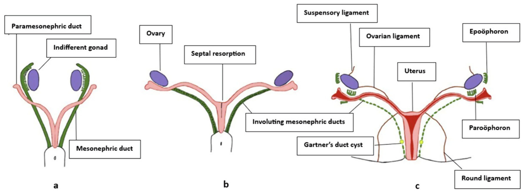
↓  Figure 1. Development of female internal genitalia. (a) Undifferentiated gonads are present with Mullerian ducts lateral to mesonephric ducts cranially and medial caudally. (b) In females, mesonephric ducts regress as Mullerian ducts fuse and the septum is resorbed. (c) Uterus, cervix, and upper vagina, with mesonephric remnants such as the epoophoron, paroophoron, and Gartner’s duct cysts [5].
Figure 1.
↓  Figure 2. Transvaginal ultrasound. (a) View of two cervices (yellow arrows). (b) Transverse view of normal uterus.
Figure 2.
↓  Figure 3. MRI of normal sagittal view of uterus. MRI: magnetic resonance imaging.
Figure 3.
↓  Figure 4. MRI of double cervix at external os (yellow arrows). MRI: magnetic resonance imaging.
Figure 4.
↓  Figure 5. MRI of vaginal septum (yellow arrows). MRI: magnetic resonance imaging.
Figure 5.
↓  Figure 6. MRI showing duplication of vagina (yellow arrows). MRI: magnetic resonance imaging.
Figure 6.
↓  Figure 7. Diagram of genital tract of patient (U0, C2, V1 using ESHRE classification). ESHRE: the European Society of Human Reproduction and Embryology.
Figure 7.