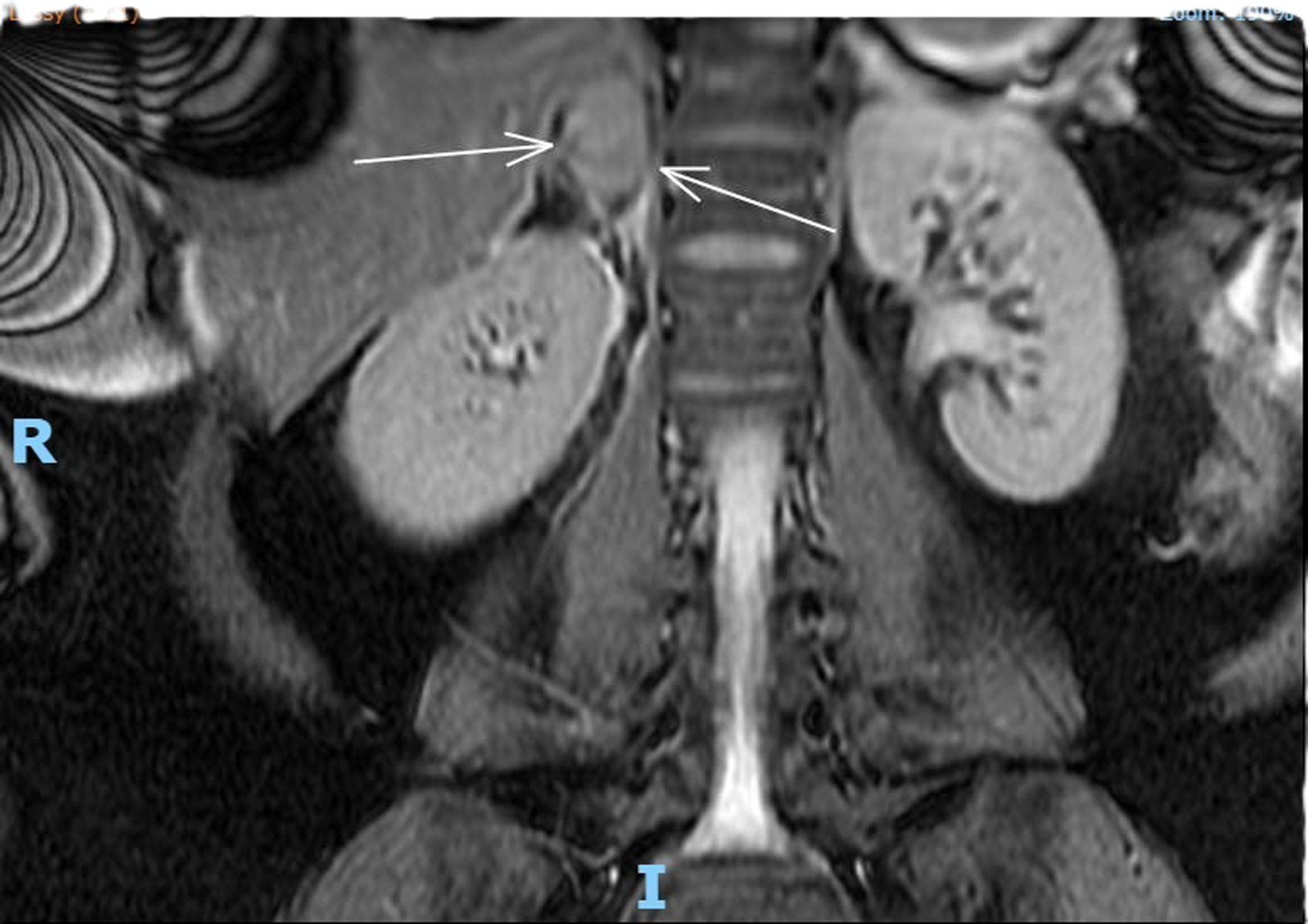
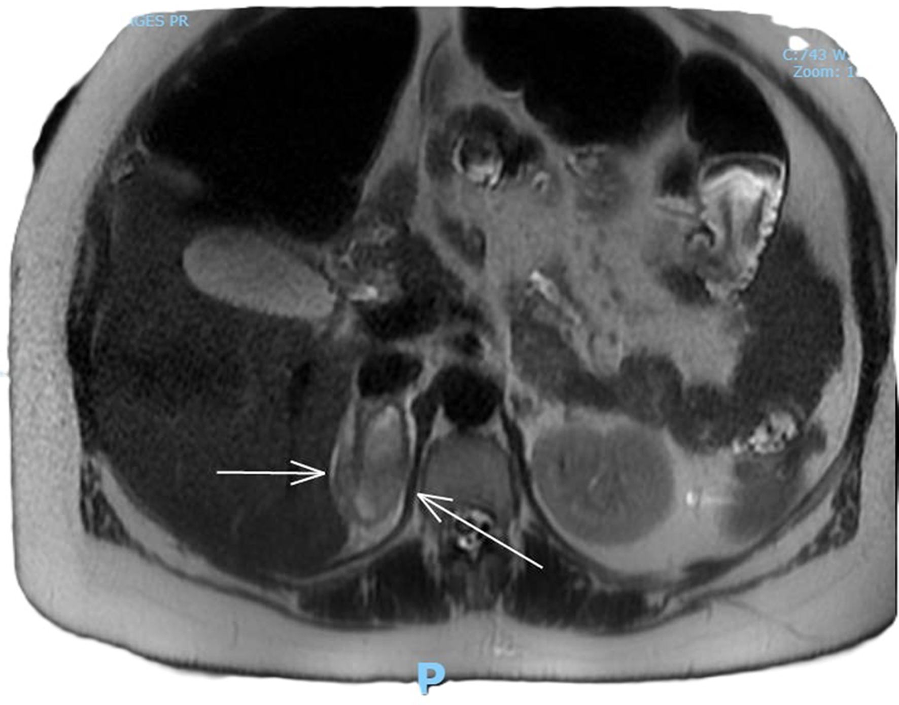
↓ Figure 1. MRI coronal plane, T2-weighted
sequence demonstrating an area of heterogeneous high T2 signal density. An arrow points to enlarged
adrenal gland consistent with adrenal hemorrhage.
| Journal of Clinical Gynecology and Obstetrics, ISSN 1927-1271 print, 1927-128X online, Open Access |
| Article copyright, the authors; Journal compilation copyright, J Clin Gynecol Obstet and Elmer Press Inc |
| Journal website https://jcgo.elmerpub.com |
Case Report
Volume 14, Number 4, December 2025, pages 189-194
Spontaneous Adrenal Hemorrhage in Pregnancy
Figures