Journal of Clinical Gynecology and Obstetrics, ISSN 1927-1271 print, 1927-128X online, Open Access
Article copyright, the authors; Journal compilation copyright, J Clin Gynecol Obstet and Elmer Press Inc
Journal website https://jcgo.elmerpub.com

Case Report

Volume 000, Number 000, March 2025, pages 000-000


A Tubo-Ovarian Abscess Caused by Salmonella enterica

Figures

Figure 1.
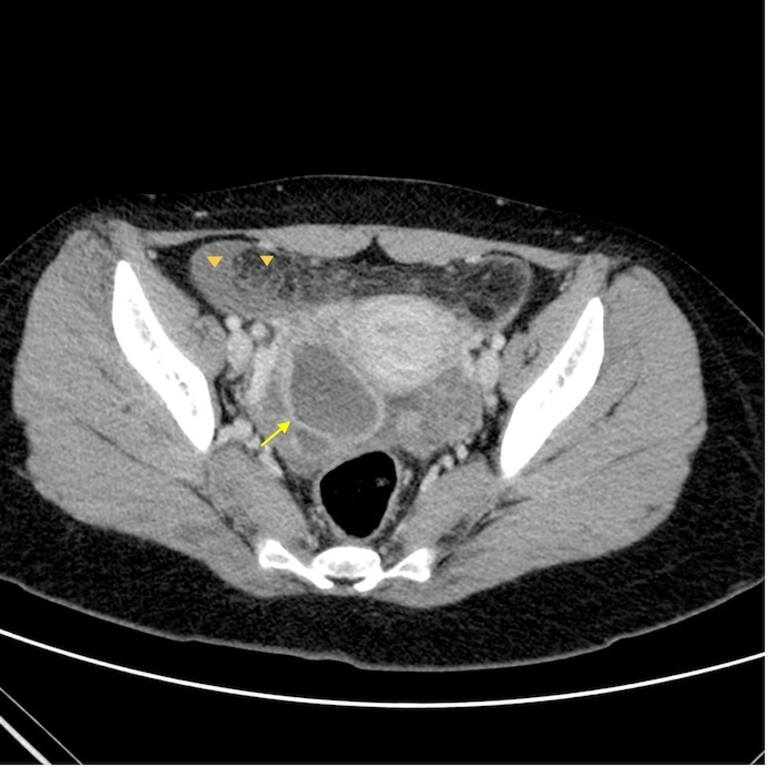
Figure 1. Axial post-contrast computed tomography (CT) scan of the abdomen shows a cystic lesion in the right adnexa (arrow) with a thick enhancing wall. Fat stranding and free fluid are also present (arrowheads).
Figure 2.
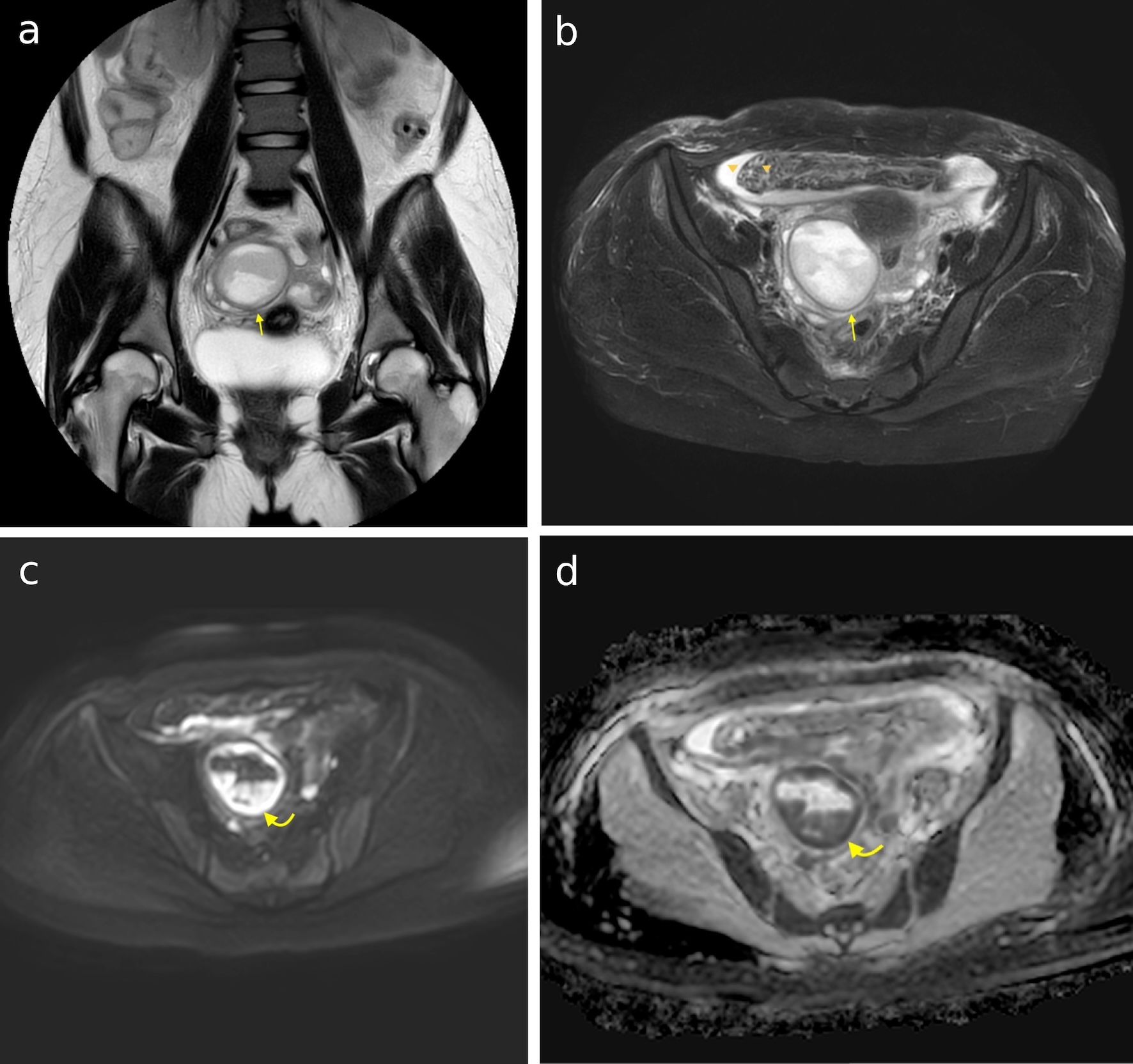
Figure 2. (a) Coronal T2-weighted image of the abdomen shows a highly heterogeneous complex cystic lesion in the right side of the pelvis (arrow). (b) Axial T2-weighted fat-saturated image shows a large complex cystic lesion in the right side of the pelvis (arrow), displacing the right adnexa inferiorly. Fat stranding and free fluid are also present (arrowheads). (c) Axial DWI image at b = 1,000 s/mm2, showing areas of high signal intensity within the lesion (curved arrow). (d) Same areas depict low signal in the ADC map corresponding to areas of restricted diffusion (curved arrow). DWI: diffusion-weighted imaging; ADC: apparent diffusion coefficient.