Journal of Clinical Gynecology and Obstetrics, ISSN 1927-1271 print, 1927-128X online, Open Access
Article copyright, the authors; Journal compilation copyright, J Clin Gynecol Obstet and Elmer Press Inc
Journal website https://jcgo.elmerpub.com

Review

Volume 14, Number 1, January 2025, pages 1-13


Abnormal Uterine Bleeding: Saudi Guideline

Figure

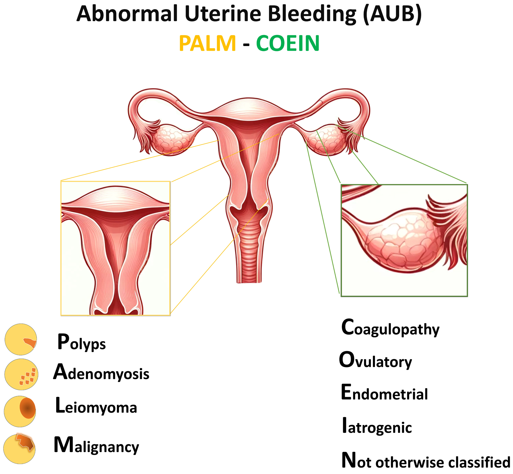
Figure 1.
Figure 1. Abnormal uterine bleeding (AUB) PALM-COEIN classification. P: polyp, A: adenomyosis, L: leiomyoma, M: malignancy and hyperplasia, C: coagulopathy, O: ovulatory dysfunction, E: endometrial disorders, I: iatrogenic, N: not otherwise classified.

Tables

Table 1. Patient History and Examination for Suspected AUB
 
Patient history
The clinician should obtain a detailed history from a patient who presented with complaints related to menstruation [15, 16]:
1) Menstrual history, including age at menarche, last menstrual period, and intermenstrual and postcoital bleeding. Additionally, the frequency, regularity, duration, and the volume of flow of menses should be recorded:
a) Frequency can be described as frequent (less than 24 days), normal (24 to 38 days), or infrequent (greater than 38 days).
b) Regularity can be described as absent, regular (with a variation of ± 2 to 7 days), or irregular (variation greater than 20 days).
c) The duration can be described as prolonged (greater than 8 days), normal (approximately 4 to 8 days), or shortened (less than 4 days).
d) The volume of flow can be described as heavy (greater than 80 mL), normal (5 to 80 mL), or light (less than 5 mL of blood loss)a.
2) Sexual and reproductive history: obstetrical history, including the number of pregnancies and mode of delivery, fertility desire and subfertility, current contraception, possibility of pregnancy, history of STIs, history of Pap smear, history of vaccination, and any previous diagnostic management (i.e., endometrial biopsy, etc.).
3) Systemic symptoms such as any history of weight loss, pain, discharge, bowel or bladder symptoms, symptoms or history of bleeding disorders, symptoms or history of endocrine disorders.
4) Symptoms of anemia: headache, palpitations, shortness of breath, dizziness, fatigue, and pica.
5) Associated symptoms: fever, chills, increasing abdominal girth, pelvic pressure or pain, bowel or bladder dysfunction, vaginal discharge, or odor.
6) Symptoms associated with a systemic cause for AUB: overweight, obesity, PCOS, hypothyroidism, hyperprolactinemia, hypothalamic or adrenal disorder.
7) Chronic medical illness: inherited bleeding disorders (coagulopathy, blood dyscrasias, and platelet functional disorders), systemic lupus erythematosus or other connective tissue diseases, liver disease, renal disease, and cardiovascular disease.
8) Current medication intake: hormonal contraceptives, anticoagulants, SSRIs, antipsychotics, tamoxifen, and herbals (e.g., Ginseng).
9) Family history including questions concerning coagulopathies, malignancy, and endocrine disorders.
10) Social history including tobacco, alcohol, and drug uses; occupation; the impact of symptoms on quality of life.
11) Surgical history.
Physical exam
aExact volume measurements are difficult to determine outside research settings; therefore, detailed questions regarding frequency of sanitary product changes during each day, passage and size of any clots, need to change sanitary products during the night, and a “flooding” sensation is important. AUB: abnormal uterine bleeding; BMI: body mass index; PCOS: polycystic ovary syndrome; SSRIs: selective serotonin reuptake inhibitors; STI: sexually transmitted infection.
Attention should be given to the general well-being of the patient. The physical exam should include [15, 16]:
1) Taking vital signs, including pulse, blood pressure, weight, and BMI. Orthostatics as clinically indicated.
2) Examination for pallor looking for signs of skin or mucosal pallor.
3) Examination of the thyroid for enlargement or tenderness (signs of endocrine disorders).
4) Checking for excessive hair growth, clitoromegaly, and acne (signs of hyperandrogenism).
5) Checking for moon facies, abnormal fat distribution, and striae (possible indications of Cushing syndrome).
6) Examination for bruising or petechiae (signs of coagulopathies).
7) Examination of the uterus and adnexal structures to check the uterine size (normal or enlarged), contour (smooth and symmetrical or irregular), consistency (firm or soft), mobility and tenderness, and presence of adnexal masses.
8) Examination of the abdomen for tenderness, distension, striae, palpable mass, and hepatomegaly.
9) Examination of the pelvis with speculum to check:
a) The vulva for trauma or skin lesions.
b) The vagina for vaginitis, trauma, and ulcerative lesions.
c) The cervix for ectropion, cervicitis, polyps, or focal lesions.
d) The anus for anal fissures, hemorrhoids, inflammatory bowel disease, and cancers.
e) The urethra for urethritis or caruncle.
f) Pap smear, if indicated.
g) A screening for STIs such as for gonorrhea and chlamydia and wet prep if indicated.
h) Endometrial biopsy, if indicated.
10) Examination of the rectum if bleeding from rectum suspected or risk of concomitant pathology.


Table 2. Laboratory Tests for Evaluating Patients With AUBa
 
EvaluationDetailed laboratory tests
aAdapted from ACOG 2013 [17]. AUB: abnormal uterine bleeding; DHEA-S: dehydroepiandrosterone sulfate; FSH: follicle-stimulating hormone; LH: luteinizing hormone; PCOS: polycystic ovary syndrome.
Initial laboratory testingPatients with no homeostasis disorders:
1) Complete blood count
2) Serum ferritin
3) Sex hormones: LH, FSH, prolactin
4) Thyroid-stimulating hormone
5) Pregnancy test
Additional tests to considerPatients with homeostasis disorders:
1) Partial thromboplastin time
2) Prothrombin time
3) Activated partial thromboplastin time
4) Fibrinogen level
Patients with von Willebrand disease:
1) von Willebrand factor antigen
2) Ristocetin cofactor assay
3) Factor VIII
Patient’s findings suggestive of PCOS:
1) Testosterone (free/total)
2) DHEA-S


Table 3. Imaging Modalities to Evaluate AUB
 
Imaging modalityAdvantages
AUB: abnormal uterine bleeding; HSG: hysterosalpingography; MRI: magnetic resonance imaging; SIS: saline infusion sonography; 3D: three-dimensional; TVS: transvaginal sonography; TVUS: transvaginal ultrasound.
TVUSSimple, affordable, widely available, and is the gold standard in assessing the endometrium, myometrium, and the cervix. Whether an intracavitary lesion (polyp), endometrial texture changes (hyperplasia), or myometrial mass (fibroid) all can be captured with relatively high accuracy with TVUS. 3D TVUS can be of additional benefit.
Color flow DopplerIt is helpful to distinguish adenomyosis from fibroids, and polyps from submucosal leiomyoma. More importantly, it is useful for the detection of arteriovenous malformations of the uterus.
SISIt has a comparable sensitivity to hysteroscopy for the diagnosis of intracavitary polyps and submucosal myomas.
MRIThis modality is effective at distinguishing adenomyomas from leiomyomas (class II-1). It is superior to TVS, SIS, and hysteroscopy for measuring the myometrial extent of submucous leiomyomas. It can miss a polyp.
HSGIt is less accurate than hysteroscopy for cavity evaluation with suboptimal sensitivity and specificity.


Table 4. Office Special Investigations for AUB Evaluation
 
InvestigationAdvantages
AUB: abnormal uterine bleeding.
Office endometrial biopsyIt is a relatively quick and cost-effective way to sample the endometrium in women above the age of 45 with high accuracy in detecting endometrial hyperplasia or malignancy.
Pap smearAbnormal Pap results have been documented in AUB caused by cervical neoplastic lesions.
Office hysteroscopyIts convenient establishment in an office allows instant identification of endometrial lesions with immediate action of removal or getting a biopsy. A trained gynecologist is required.