Journal of Clinical Gynecology and Obstetrics, ISSN 1927-1271 print, 1927-128X online, Open Access
Article copyright, the authors; Journal compilation copyright, J Clin Gynecol Obstet and Elmer Press Inc
Journal website https://jcgo.elmerpub.com

Case Report

Volume 14, Number 1, January 2025, pages 25-30


Spontaneous Subarachnoid Hemorrhage After Cesarean Section

Figures

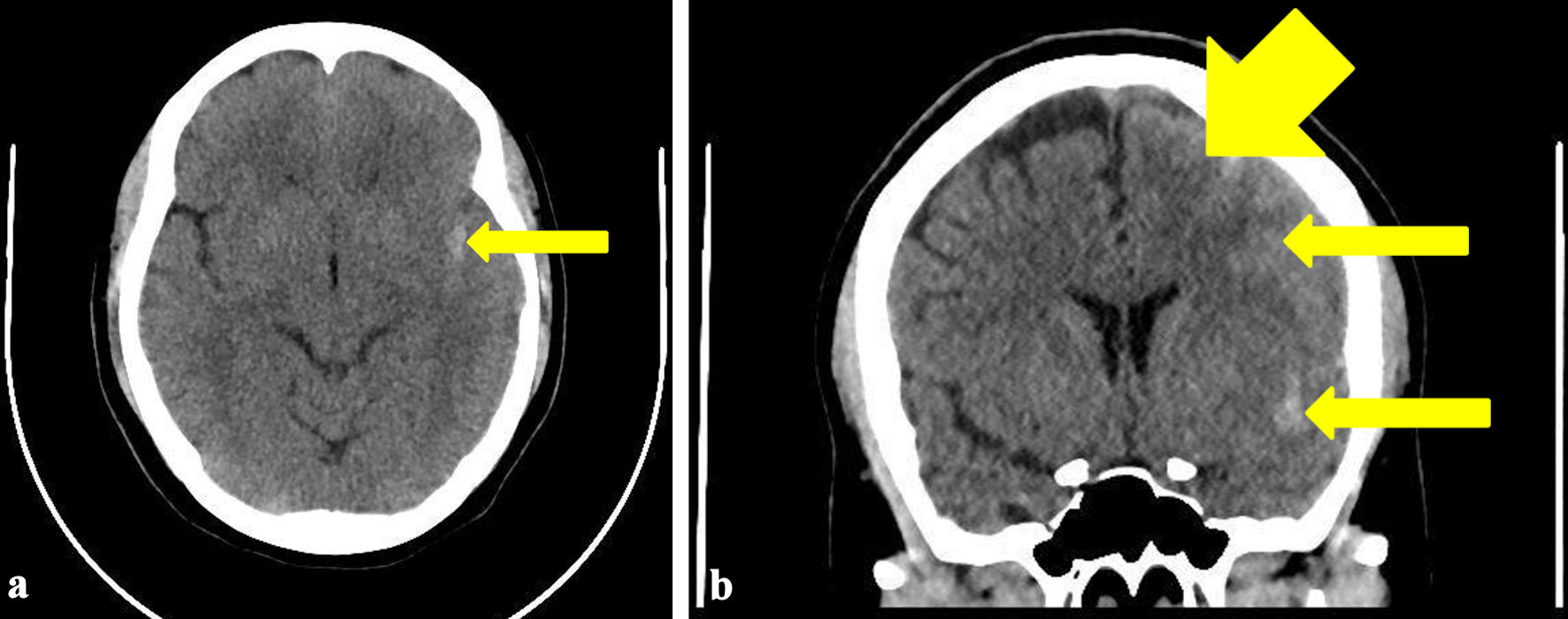
↓  Figure 1. Transverse view (a) and coronal view (b) of the CT brain showing hyper-densities within left sylvian fissure (arrow) and over left anterior frontal lobe (arrows) consistent with widespread acute subarachnoid hemorrhage. Mild diffuse edema is observed in the left cerebral hemisphere. CT: computed tomography.
Figure 1.
↓  Figure 2. CT angiography of the brain showing normal appearance of the circle of Willis. Normal angiographic appearance of both vertebral arteries, basilar artery and posterior cerebral arteries (arrow). CT: computed tomography.
Figure 2.