| Journal of Clinical Gynecology and Obstetrics, ISSN 1927-1271 print, 1927-128X online, Open Access |
| Article copyright, the authors; Journal compilation copyright, J Clin Gynecol Obstet and Elmer Press Inc |
| Journal website https://jcgo.elmerpub.com |
Case Report
Volume 14, Number 4, December 2025, pages 195-198

Spontaneous Hemoperitoneum in Advanced Maternal Age Pregnancy
Elizabeth Stilesa, Linda Zambrano Guevaraa, Andrew Rubensteina, b
aDepartment of Obstetrics and Gynecology, NYU Langone Health, New York, NY,
USA
bCorresponding Author: Andrew Rubenstein, Department of Obstetrics and
Gynecology, NYU Langone Health, New York, NY 10022, USA
Manuscript submitted September 4, 2025, accepted November 21, 2025, published online December 11,
2025
Short title: Spontaneous Hemoperitoneum in Pregnancy
doi:
https://doi.org/10.14740/jcgo1544
| Abstract | ▴Top |
Spontaneous hemoperitoneum in pregnancy (SHiP) is a rare but serious condition. Risk factors include endometriosis, in vitro fertilization (IVF), prior pelvic surgeries, and advanced maternal age (AMA). We report a 41-year-old female at 36 weeks’ gestation who presented with severe abdominal pain and non-reassuring fetal heart rate. Urgent cesarean delivery (CD) revealed hemoperitoneum. Clear amniotic fluid pointed to an extra-uterine source: bleeding from the posterior uterine serosa. While prompt surgical intervention in this case ensured maternal-fetal safety, the potential value of imaging to improve earlier recognition and diagnosis is discussed. This case underscores the need to consider SHiP in pregnancies lacking other known risk factors and presenting with unexplained abdominal symptoms.
Keywords: Advanced maternal age; Endometriosis; IVF; Ultrasound imaging
| Introduction | ▴Top |
Spontaneous hemoperitoneum in pregnancy (SHiP) is a rare but life-threatening obstetric complication defined by intraperitoneal bleeding without an external traumatic cause. SHiP carries significant risks to maternal-fetal health, with reported perinatal mortality rates reaching up to 31% in some reviews [1]. Clinical presentation ranges from nonspecific abdominal pain to signs of hypovolemic shock and fetal distress [2, 3]. Because symptoms often mimic more common obstetric conditions, diagnosis may be delayed, likely contributing to poor outcomes [4, 5].
Most documented cases of SHiP involve underlying risk factors such as endometriosis, in vitro fertilization (IVF), and prior pelvic surgery [6-9]. In particular, endometriosis is thought to contribute to vascular fragility through chronic inflammation and decidualization of ectopic implants during pregnancy [2, 10, 11]. IVF-related hormonal changes and pelvic adhesions have also been implicated in increasing vascular rupture risk [6, 12]. Similarly, post-surgical intrabdominal inflammation is hypothesized to weaken vessel walls and promote adhesion formation [9]. However, a subset of SHiP cases have been reported in patients lacking these conventional risk factors [3, 13, 14]. These findings underscore that SHiP can occur even in patients without a notable medical or surgical history, reinforcing the need for clinical vigilance.
In this case report, we describe a patient presenting with SHiP whose only identifiable risk factor was advanced maternal age (AMA). By contributing to the limited pool of contemporary reports, this case highlights the importance of considering SHiP in the differential diagnosis of acute abdomen during pregnancy in patients lacking more well-documented risk factors.
| Case Report | ▴Top |
A 41-year-old Asian G2P0010 female at 36 weeks’ gestation presented to the obstetrical triage of a large, urban academic hospital with severe (rated as 10 out of 10) abdominal pain starting approximately 6 h prior. She was unable to void for several hours and had consumed a “heavy” meal about 1 h prior to presentation. She reported uterine contractions occurring approximately every 6 min. She endorsed ongoing fetal movements, and denied vaginal bleeding or leakage of fluid.
Her pregnancy was spontaneously conceived and managed with routine prenatal care initiated during the first trimester. Her pregnancy was complicated by AMA, low-lying placenta (resolved), nausea and vomiting (taking doxylamine-pyridoxine and ondansetron), and anxiety (started sertraline 50 mg daily in the third trimester). She was also taking aspirin (81 mg daily starting at 12 weeks’ gestation) for preeclampsia prophylaxis. Her medical history was significant for obesity (pregravid body mass index (BMI) of 34), gastroesophageal reflux disease (on famotidine), migraines, and a uterine fibroid. She had undergone a dilation and curettage following a spontaneous abortion 1 year prior.
On examination, the abdomen was soft and non-tender. Pelvic examination revealed cervical dilation of 1 cm, 60% effacement, and a fetal station of -3. Bedside ultrasound showed footling breech presentation and umbilical cord close to cervix. Evaluation for free fluid in the abdomen was not performed. Fetal heart rate monitoring revealed moderate variability with recurrent late and deep variable decelerations. Labs were notable for hemoglobin of 9.1 g/dL (baseline 10.2 g/dL from 23 days prior). Given breech presentation in addition to this non-reassuring fetal status in the setting of preterm labor and concern for placental abruption, an urgent cesarean delivery (CD) was recommended. Initially, the patient declined CD despite counseling on increased risk of fetal death, anoxia, cerebral palsy, and breech delivery with head entrapment or cord prolapse. However, she consented approximately 1 h later and was taken to the operating room. She declined regional anesthesia despite counseling and opted for general anesthesia.
Upon entering the abdominal cavity and prior to uterine incision, approximately 1 to 1.5 L of blood were noted, prompting involvement of the obstetric hemorrhage team. During hysterotomy and delivery of the infant, amniotic fluid was noted to be clear. The infant was delivered without complications, weighing 5 pounds 7.1 ounces, with APGAR scores of 6 and 8 at 1 and 5 min, respectively. The placenta appeared grossly normal. Umbilical cord blood gas analysis revealed an arterial pH of 7.14 with base excess of -8.7 mmol/L, and a venous pH of 7.18 with base excess of -8.0 mmol/L. These results are consistent with moderate fetal acidemia, without evidence of severe metabolic acidosis, and adequate sampling. The patient received 2 units of packed red blood cells, 2 units of fresh frozen plasma, 1 unit of platelets, and 1 L of lactated Ringer’s solution for resuscitation.
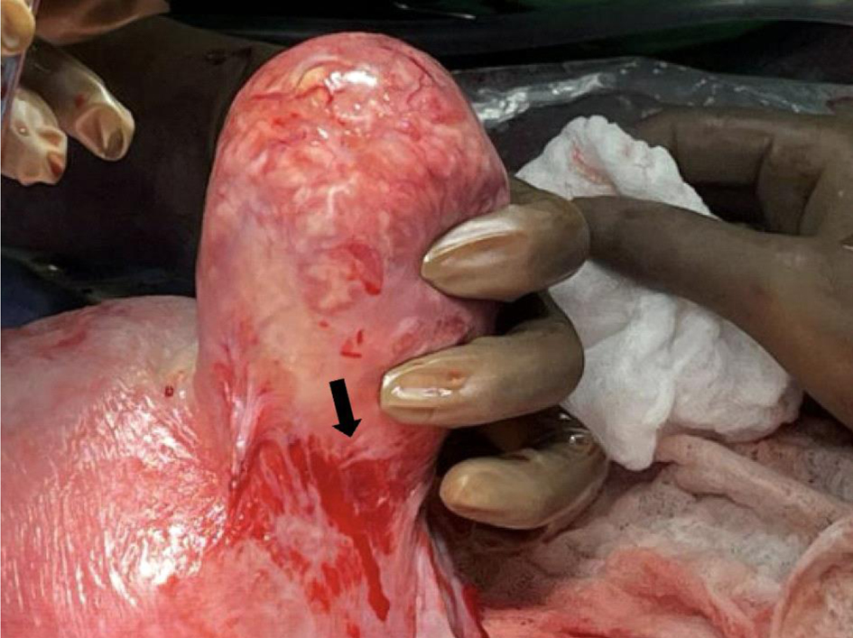
The uterus was then exteriorized and hysterotomy was closed (Fig. 1). Several large clots posterior to the uterus and an approximately 2 cm area of active bleeding on the posterior uterine serosa and superficial myometrium were noted (Fig. 2). The uterus was fully intact with no signs of uterine rupture, arteriovenous malformations, varices, or endometriotic implants noted. A full survey of the abdominal cavity was performed, and the adnexa, bowel, peritoneum, and mesentery all appeared grossly normal. Hemostasis at the site of serosal bleeding was achieved using 0-Vicryl running locked sutures. A separate 8 cm right subserosal fibroid was identified with minimal oozing (no substantial contribution to the hemoperitoneum), likely from disruption during uterine manipulation (Fig. 3). This bleeding was controlled using 0-monocryl sutures. Total measured blood loss was 2 L. Tranexamic acid, methergine, and carboprost tromethamine were administered.
 Click for large image |
Figure 1. The exteriorized uterus demonstrating a repaired 2 cm site of bleeding (arrow) and 8 cm subserosal fibroid (star). |
 Click for large image |
Figure 2. Posterior uterine serosa and superficial myometrium with 2 cm site of bleeding after suture repair (arrow). |
 Click for large image |
Figure 3. The 8 cm right subserosal fibroid with minimal bleeding (arrow). |
The patient received redosing of cefazolin intraoperatively for infection prophylaxis. Postoperatively, she developed acute blood loss anemia but declined transfusion of a third unit of packed red blood cells, instead receiving intravenous iron. Her Foley catheter was removed on postoperative day (POD) 1. On POD 2, she was diagnosed with an ileus, which was managed with nasogastric tube decompression until clinical improvement on POD 3. Pain control was achieved using patient-controlled analgesia with intravenous hydromorphone, alongside supportive medications including ondansetron and famotidine as needed. She was discharged home on POD 5 with a 6-week course of low molecular weight heparin for venous thromboembolism prophylaxis (Royal College of Obstetricians and Gynaecologists score 5). Both mother and infant were scheduled for routine postpartum and pediatric follow-up, respectively.
| Discussion | ▴Top |
SHiP is a rare but critical condition that can present with non-specific abdominal complaints, often mimicking more common obstetrical or gastrointestinal issues [4]. Common obstetric causes of acute abdominal pain include placental abruption, uterine rupture, labor contractions, and round ligament pain, while non-obstetric causes include appendicitis, cholecystitis, bowel obstruction, renal colic, and pancreatitis. In contrast, SHiP often presents as sudden, severe, nonlocalized abdominal pain in the absence of vaginal bleeding or uterine tenderness. SHiP may occur even when vital signs and hemoglobin are initially near normal, delaying its inclusion in a clinician’s differential diagnosis. In this case, the patient presented with intense abdominal pain but no vaginal bleeding or severe anemia. While uterine contractions and non-reassuring fetal heart rate tracing initially raised concern for placental abruption, the placenta appeared grossly normal. The discovery of 1 to 1.5 L of hemoperitoneum with clear amniotic fluid at the time of CD indicated an extra-uterine etiology. Thus, it is likely that maternal blood loss leading to uteroplacental hypoperfusion as well as uterine irritability contributed to fetal heart rate abnormalities.
Earlier use of abdominal imaging such as ultrasound, magnetic resonance imaging (MRI), or computed tomography angiography (CTA) may have aided in preoperative recognition of intra-abdominal bleeding. Several case reports emphasize the diagnostic value of imaging when SHiP is suspected, especially in patients without vaginal bleeding [2, 7]. In addition to gathering a detailed medical history (including risk factors like endometriosis, IVF, and prior pelvic surgeries), performing a thorough physical exam, and monitoring hemoglobin levels, physicians should consider imaging for free intra-abdominal fluid if maternal and fetal conditions are stable [8]. Given the severity of abdominal pain, moderate anemia slightly lower than baseline, and fetal distress, focused assessment with sonography for trauma (FAST) may have been especially useful in this urgent presentation and should be prioritized as a first-line modality in acute obstetrical triage.
FAST offers rapid bedside assessment for intra-abdominal free fluid and can be performed without moving the patient or exposing the fetus to radiation, making it most practical in emergency obstetric settings. MRI provides superior soft-tissue contrast and can better localize vascular or endometriotic sources of bleeding, but is limited by availability, scan time, and patient stability. CTA remains the most sensitive for active extravasation or vascular injury but is generally reserved for hemodynamically stable patients with known but acceptable risk of radiation and contrast exposure. Therefore, a pragmatic diagnostic approach in suspected SHiP may begin with bedside FAST for rapid triage, followed by MRI or CTA only if the patient is stable and initial findings are inconclusive. In cases where maternal or fetal compromise persists despite uncertainty in diagnosis, expedited surgical exploration is recommended.
This case also underscores the importance of considering extra-uterine bleeding sources when amniotic fluid appears clear after hysterotomy. In several reports of SHiP, clear amniotic fluid helped localize the hemorrhagic source to the peritoneal rather than the intrauterine space [3, 13]. In our case, the identification of active bleeding on the posterior uterine serosa supported this differentiation. The differential diagnosis for extra-uterine sources of bleeding during pregnancy includes rupture of uterine serosal or utero-ovarian vessels, bleeding from decidualized endometriosis, vascular malformations, or avulsion injuries, none of which were visibly present in this patient [1, 12]. However, it is possible that the patient’s history of dilation and curettage may have resulted in localized uterine scarring or altered vascular integrity not appreciated on exam or prior imaging, predisposing to abnormal vessel remodeling in subsequent pregnancy. Elevated intra-abdominal pressure and chronic low-grade inflammation associated with obesity could also result in endothelial dysfunction and impaired wound healing, theoretically heightening risk for vessel rupture during pregnancy.
Though most published cases link SHiP to risk factors such as endometriosis, IVF, or prior pelvic surgery, our patient’s only risk factor was AMA, consistent with a small but meaningful number of SHiP cases reported in similarly minimal-risk populations. The underlying mechanism linking AMA to increased SHiP risk is unknown, but may be confounded by higher likelihood of IVF and prior pelvic surgeries in this population [9]. While low-dose aspirin has been associated with increased risk of intra- and postpartum bleeding, the absolute increase in blood loss is small for most patients and thus unlikely to be the main driver of this patient’s massive hemorrhage [15, 16].
The presence of a bleeding subserosal fibroid adds complexity to the clinical interpretation. While fibroids are not a widely documented cause of SHiP, manipulation of the uterus during delivery may have disrupted vascular supply to the fibroid or surrounding tissue. Suspicion of superficial vascular rupture on the uterine surface in the absence of endometriosis has been documented in other cases, raising the possibility that even non-endometriotic lesions like fibroids can contribute to vascular compromise under mechanical stress [3, 11, 17].
Triage in cases like this should involve rapid maternal-fetal assessment, including fetal heart monitoring, laboratory evaluation of hemoglobin levels, and consideration of rapid imaging modalities like FAST [8]. Early multidisciplinary involvement, including obstetrics, emergency medicine, anesthesiology, and transfusion medicine, is essential. Given signs of fetal distress, the decision for urgent CD was appropriate in this case. Timely recognition and management coupled with patient education and shared decision-making given the patient’s initial reluctance to proceed with CD ultimately promoted favorable maternal and neonatal outcomes.
Learning points
Providers should maintain a high index of suspicion for SHiP even in the absence of more common risk factors. Early imaging and multidisciplinary involvement are critical when pregnant patients present with acute abdominal pain and non-reassuring fetal heart rate. Prompt surgical intervention may be lifesaving in cases of intra-abdominal bleeding during pregnancy, underscoring the importance of timely diagnostic and operative decision-making.
Acknowledgments
None to declare.
Financial Disclosure
The authors declare no financial disclosures related to this work.
Conflict of Interest
The authors declare no conflict of interest related to this work.
Informed Consent
Signed written consent for publication was obtained from the patient and kept on file.
Author Contributions
Elizabeth Stiles: data curation, investigation, and writing – original draft. Linda Zambrano Guevara and Andrew Rubenstein: resources, clinical insights, and writing – review and editing.
Data Availability
Any inquiries regarding supporting data availability of this study should be directed to the corresponding author.
| References | ▴Top |
- Ruxandra-Patricia N, Ciobanu AM, Gica C, Demetrian M, Cimpoca-Raptis
BA, Peltecu G, et al. Spontaneous hemoperitoneum in pregnancy. Ro Med J.
2022;69(S2):23-26.
doi - Sim Y, Kim J, Jeong Y, Rheu C, Chae H. Spontaneous hemoperitoneum in
pregnancy (SHiP) complicated by endometriosis: A case report. Obstet Gynecol Rep.
2020;4(2).
doi - Owie E. Incidental hemoperitoneum from ruptured superficial uterine
veins in twin pregnancy. Trop J Obstet Gynaecol. 2018;35(3).
doi - Mamah J, Thomas M, Rafi J. Spontaneous hemoperitoneum in pregnancy:
masquerading as acute appendicitis. Cureus. 2023;15(10):e47040.
doi pubmed - Dhamecha R, Pajai S, Bhasin T. Acute abdomen in pregnancy: a
comprehensive review of diagnosis and management. Cureus. 2023;15(6):e40679.
doi pubmed - Brosens IA, Lier MC, Mijatovic V, Habiba M, Benagiano G. Severe
spontaneous hemoperitoneum in pregnancy may be linked to in vitro fertilization in patients with
endometriosis: a systematic review. Fertil Steril. 2016;106(3):692-703.
doi pubmed - Vuong ADB, Pham TH, Nguyen XT, Trinh NB, Nguyen PN, Ho QN.
Spontaneous hemoperitoneum in the second and third trimester of pregnancy: two uncommon case
reports at Tu Du Hospital, in Vietnam and a literature review. Int J Emerg Med.
2023;16(1):26.
doi pubmed - Schreurs AMF, Overtoom EM, de Boer MA, van der Houwen LEE, Lier MCI,
van den Akker T, Cornette J, et al. Spontaneous haemoperitoneum in pregnancy: Nationwide
surveillance and Delphi audit system. BJOG. 2023;130(13):1620-1628.
doi pubmed - Mazzocco MI, Donati S, Maraschini A, Corsi E, Colciago E, Guelfi F,
Cetin I. Spontaneous hemoperitoneum in pregnancy: Italian prospective population-based cohort
study. Acta Obstet Gynecol Scand. 2022;101(11):1220-1226.
doi pubmed - Katorza E, Soriano D, Stockheim D, Mashiach R, Zolti M, Seidman DS,
Schiff E, et al. Severe intraabdominal bleeding caused by endometriotic lesions during the third
trimester of pregnancy. Am J Obstet Gynecol. 2007;197(5):501.e501-504.
doi pubmed - Kato M, Miyazaki Y, Kawamura H, Orisaka M, Kurokawa T, Yoshida Y. A
41-year-old woman with spontaneous hemoperitoneum in pregnancy at 28 weeks.
Am J Case Rep. 2023;24:e939330.
doi pubmed - Lier MCI, Malik RF, Ket JCF, Lambalk CB, Brosens IA, Mijatovic V.
Spontaneous hemoperitoneum in pregnancy (SHiP) and endometriosis - A systematic review of the
recent literature. Eur J Obstet Gynecol Reprod Biol. 2017;219:57-65.
doi pubmed - Xu Y, Zhou Y, Xie J, Yin X, Zhang X. Intraperitoneal hemorrhage
during pregnancy and parturition: Case reports and literature review. Medicine (Baltimore).
2019;98(35):e16300.
doi pubmed - Yang L, Liu N, Long Y. Intra-abdominal hemorrhage during pregnancy:
four case reports. World J Clin Cases. 2020;8(14):3074-3081.
doi pubmed - Jiang Y, Chen Z, Chen Y, Wei L, Gao P, Zhang J, Zhou X, et al.
Low-dose asprin use during pregnancy may be a potential risk for postpartum hemorrhage and
increased blood loss: a systematic review and meta-analysis. Am J Obstet Gynecol
MFM. 2023;5(4):100878.
doi pubmed - Hastie R, Tong S, Wikstrom AK, Sandstrom A, Hesselman S, Bergman L.
Aspirin use during pregnancy and the risk of bleeding complications: a Swedish population-based
cohort study. Am J Obstet Gynecol. 2021;224(1):95.e1-e12.
doi pubmed - Akriti G, Uttara G, Anand G. The case series of spontaneous
hemoperitoneum in third trimester pregnancy. Asian J Med Sci.
2022;13(9):268-270.
doi
This
article is distributed under the terms of the Creative Commons Attribution Non-Commercial 4.0
International License, which permits unrestricted non-commercial use, distribution, and
reproduction in any medium, provided the original work is properly cited.
Journal
of Clinical Gynecology and Obstetrics is published by Elmer Press Inc.